|
|
|
| Knowledge-guided infarct segmentation of ischemic stroke |
Zhengyu GU1( ),Feifei LAI2,Chen GENG3,Ximing WANG4,Yakang DAI1,3,*() ),Feifei LAI2,Chen GENG3,Ximing WANG4,Yakang DAI1,3,*() |
1. School of Medical Imaging, Xuzhou Medical University, Xuzhou 221004, China
2. Department of Radiology, Wuxi Mental Health Center, Wuxi 214151, China
3. Suzhou Institute of Biomedical Engineering and Technology, Chinese Academy of Sciences, Suzhou 215163, China
4. Department of Radiology, The First Affiliated Hospital of Soochow University, Suzhou 215006, China |
|
|
|
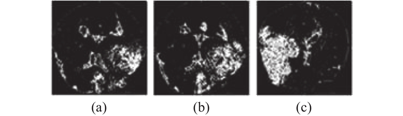
Abstract Ischemic stroke infarcts show low-density features on imaging. Based on threshold segmentation and weighted filtering for the features, an infarct probability map generation method was proposed. The low-density region was identified by threshold segmentation with adaptive parameters, and the infarct probability map was obtained by calculating the binary map through the filter with customized weights at multiscale. The weights of the low-probability regions were reduced when the probability map guided the net parameters learning, thus improving the segmentation accuracy of the proposed method. The probability map was used to guide U-Net, DRINet and DeepLabV3+, the Dice coefficient was increased by 0.0466, 0.0418 and 0.0363, and the intersection over union (IoU) was increased by 0.0322, 0.0440 and 0.0356, respectively, compared to the models not guided with infarct probability maps. Statistical results show that the infarct probability map-guided network has an enhancement effect on the Dice coefficient for acute-phase data and has little effect on the segmentation results for sub-acute data. The proposed method provides a feasible solution for automatic segmentation of the acute ischemic stroke infarct.
|
|
Received: 03 December 2023
Published: 25 April 2025
|
|
|
| Fund: 国家自然科学基金资助项目(81971685,62441114);江苏省前沿引领技术基础研究项目(BK20192004);山东省自然科学基金资助项目(ZR2022QF093);苏州科技计划项目(SKY2022151);浙江省医药卫生科技计划项目(2022KY1426). |
|
Corresponding Authors:
Yakang DAI
E-mail: 2297764691@qq.com;daiyk@sibet.ac.cn
|
基于知识引导的缺血性脑卒中梗死区分割方法
针对缺血性脑卒中梗死区在医学影像上显示出低密度特征,提出基于阈值分割和加权滤波的梗死概率图生成方法. 通过自适应参数的阈值分割找出低密度区,由多尺度自定义权重的滤波器计算二值图,获得梗死概率图. 当概率图引导网络参数学习时,通过降低低概率区域的权重来提高所提方法的分割准确度. 使用梗死概率图引导U-Net、DRINet和DeepLabV3+,相比未使用梗死概率图引导的模型,Dice系数分别提升了0.0466,0.0418和0.0363,交并比(IoU)分别提升了0.0322、0.0440和0.0356. 统计结果表明,梗死概率图引导的网络对急性期数据的Dice系数有提升作用,对亚急性数据分割结果影响不大. 所提方法为自动分割急性缺血性脑卒中梗死区提供了可行方案.
关键词:
深度学习,
知识引导,
缺血性卒中,
脑卒中梗死区,
语义分割
|
|
| [21] |
YUSHKEVICH P A, GAO Y, GERIG G. ITK-SNAP: an interactive tool for semi-automatic segmentation of multi-modality biomedical images [C]// Proceedings of the 38th Annual International Conference of the IEEE Engineering in Medicine and Biology Society . Orlando: IEEE, 2016: 3342–3345.
|
|
|
| [22] |
NAJM M, KUANG H, FEDERICO A, et al Automated brain extraction from head CT and CTA images using convex optimization with shape propagation[J]. Computer Methods and Programs in Biomedicine, 2019, 176: 1- 8
doi: 10.1016/j.cmpb.2019.04.030
|
|
|
| [23] |
QIU W, YUAN J, UKWATTA E, et al Prostate segmentation: an efficient convex optimization approach with axial symmetry using 3-D TRUS and MR images[J]. IEEE Transactions on Medical Imaging, 2014, 33 (4): 947- 960
doi: 10.1109/TMI.2014.2300694
|
|
|
| [24] |
ÇIÇEK Ö, ABDULKADIR A, LIENKAMP S S, et al. 3D U-Net: learning dense volumetric segmentation from sparse annotation [C]// Medical Image Computing and Computer-Assisted Intervention . [S. l.]: Springer, 2016: 424–432.
|
|
|
| [1] |
《中国脑卒中防治报告2020》编写组 《中国脑卒中防治报告2020》概要[J]. 中国脑血管病杂志, 2022, 19 (2): 136- 144
Report on Stroke Prevention and Treatment in China Writing Group Brief report on stroke prevention and treatment in China, 2020[J]. Chinese Journal of Cerebrovascular Diseases, 2022, 19 (2): 136- 144
|
|
|
| [2] |
朱红霞, 张国柱 半夏白术天麻汤治疗脑卒中后眩晕疗效观察[J]. 安徽中医药大学学报, 2018, 37 (5): 22- 24
ZHU Hongxia, ZHANG Guozhu Clinical effect of Banxia Baizhu Tianma decoction in treatment of post-stroke vertigo[J]. Journal of Anhui University of Chinese Medicine, 2018, 37 (5): 22- 24
doi: 10.3969/j.issn.2095-7246.2018.05.007
|
|
|
| [3] |
GOYAL M, DEMCHUK A M, MENON B K, et al Randomized assessment of rapid endovascular treatment of ischemic stroke[J]. New England Journal of Medicine, 2015, 372 (11): 1019- 1030
doi: 10.1056/NEJMoa1414905
|
|
|
| [4] |
DANKBAAR J W, HORSCH A D, VAN DEN HOVEN A F, et al Prediction of clinical outcome after acute ischemic stroke: the value of repeated noncontrast computed tomography, computed tomographic angiography, and computed tomographic perfusion[J]. Stroke, 2017, 48 (9): 2593- 2596
doi: 10.1161/STROKEAHA.117.017835
|
|
|
| [5] |
王姗, 赵建华 基于CT和MRI的影像组学在缺血性脑卒中的研究进展[J]. CT理论与应用研究(中英文), 2024, 33 (1): 83- 89
WANG Shan, ZHAO Jianhua Research progress in imaging radiomics based on computed tomography and magnetic resonance in ischemic stroke[J]. Computerized Tomography Theory and Applications, 2024, 33 (1): 83- 89
|
|
|
| [6] |
POWERS W J, RABINSTEIN A A, ACKERSON T, et al Guidelines for the early management of patients with acute ischemic stroke: 2019 update to the 2018 guidelines for the early management of acute ischemic stroke[J]. Stroke, 2019, 50 (12): e344- e418
|
|
|
| [7] |
REKIK I, ALLASSONNIÈRE S, CARPENTER T K, et al Medical image analysis methods in MR/CT-imaged acute-subacute ischemic stroke lesion: segmentation, prediction and insights into dynamic evolution simulation models. A critical appraisal[J]. NeuroImage Clinical, 2012, 1 (1): 164- 178
doi: 10.1016/j.nicl.2012.10.003
|
|
|
| [8] |
KUANG H, MENON B K, QIU W Semi-automated infarct segmentation from follow-up noncontrast CT scans in patients with acute ischemic stroke[J]. Medical Physics, 2019, 46 (9): 4037- 4045
doi: 10.1002/mp.13703
|
|
|
| [9] |
KUANG H, NAJM M, MENON B K, et al. Joint segmentation of intracerebral hemorrhage and infarct from non-contrast CT images of post-treatment acute ischemic stroke patients [C]// Medical Image Computing and Computer Assisted Intervention . [S.l.]: Springer, 2018: 681–688.
|
|
|
| [10] |
KUANG H, MENON B K, QIU W. Automated infarct segmentation from follow-up non-contrast CT scans in patients with acute ischemic stroke using dense multi-path contextual generative adversarial network [C]// Medical Image Computing and Computer Assisted Intervention . [S.l.]: Springer, 2019: 856–863.
|
|
|
| [11] |
VAN VOORST H, KONDURI P R, VAN POPPEL L M, et al Unsupervised deep learning for stroke lesion segmentation on follow-up CT based on generative adversarial networks[J]. AJNR American Journal of Neuroradiology, 2022, 43 (8): 1107- 1114
doi: 10.3174/ajnr.A7582
|
|
|
| [12] |
NIE X, LIU X, YANG H, et al Fully automatic identification of post-treatment infarct lesions after endovascular therapy based on non-contrast computed tomography[J]. Neural Computing and Applications, 2023, 35 (30): 22101- 22114
doi: 10.1007/s00521-022-08094-4
|
|
|
| [13] |
LIN T Y, DOLLÁR P, GIRSHICK R, et al. Feature pyramid networks for object detection [C]// Proceedings of the IEEE Conference on Computer Vision and Pattern Recognition . Honolulu: IEEE, 2017: 936–944.
|
|
|
| [14] |
DAI J, QI H, XIONG Y, et al. Deformable convolutional networks [C]// Proceedings of the IEEE International Conference on Computer Vision . Venice: IEEE, 2017: 764–773.
|
|
|
| [15] |
VASWANI A, SHAZEER N, PARMAR N, et al. Attention is all you need [C]// Proceedings of the 31st International Conference on Neural Information Processing Systems . [S.l.]: Curran Associates Inc., 2017: 6000–6010.
|
|
|
| [16] |
LI S, ZHENG J, LI D Precise segmentation of non-enhanced computed tomography in patients with ischemic stroke based on multi-scale U-Net deep network model[J]. Computer Methods and Programs in Biomedicine, 2021, 208: 106278
doi: 10.1016/j.cmpb.2021.106278
|
|
|
| [17] |
RONNEBERGER O, FISCHER P, BROX T. U-Net: convolutional networks for biomedical image segmentation [C]// Medical Image Computing and Computer-Assisted Intervention . [S. l.]: Springer, 2015: 234–241.
|
|
|
| [18] |
CHEN L C, ZHU Y, PAPANDREOU G, et al. Encoder-decoder with atrous separable convolution for semantic image segmentation [C]// Computer Vision – ECCV 2018 . [S.l.]: Springer, 2018: 833–851.
|
|
|
| [19] |
CHEN L, BENTLEY P, MORI K, et al DRINet for medical image segmentation[J]. IEEE Transactions on Medical Imaging, 2018, 37 (11): 2453- 2462
doi: 10.1109/TMI.2018.2835303
|
|
|
| [20] |
SZEGEDY C, LIU W, JIA Y, et al. Going deeper with convolutions [C]// Proceedings of the IEEE Conference on Computer Vision and Pattern Recognition . Boston: IEEE, 2015: 1–9.
|
|
|
|
Viewed |
|
|
|
Full text
|
|
|
|
|
Abstract
|
|
|
|
|
Cited |
|
|
|
|
| |
Shared |
|
|
|
|
| |
Discussed |
|
|
|
|