|
|
|
| Lightweight recognition algorithm for OCT images of fundus lesions |
Xiao-hu HOU1,2( ),Xiao-fen JIA1,3,*(),Bai-ting ZHAO2 ),Xiao-fen JIA1,3,*(),Bai-ting ZHAO2 |
1. The First Affiliated Hospital of Anhui University of Science and Technology (Huainan First People's Hospital), Huainan 232001, China
2. Institute of Electrical and Information Engineering, Anhui University of Science and Technology, Huainan 232001, China
3. Institute of Artificial Intelligence, Anhui University of Science and Technology, Huainan 232001, China |
|
|
|
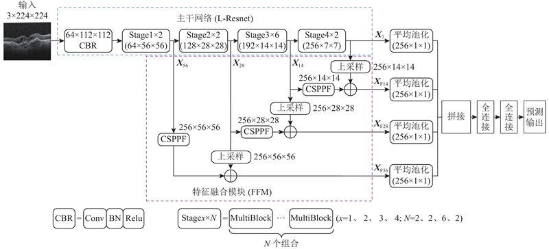
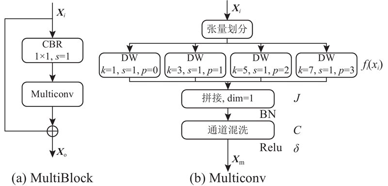
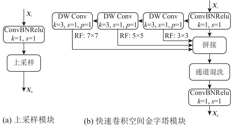
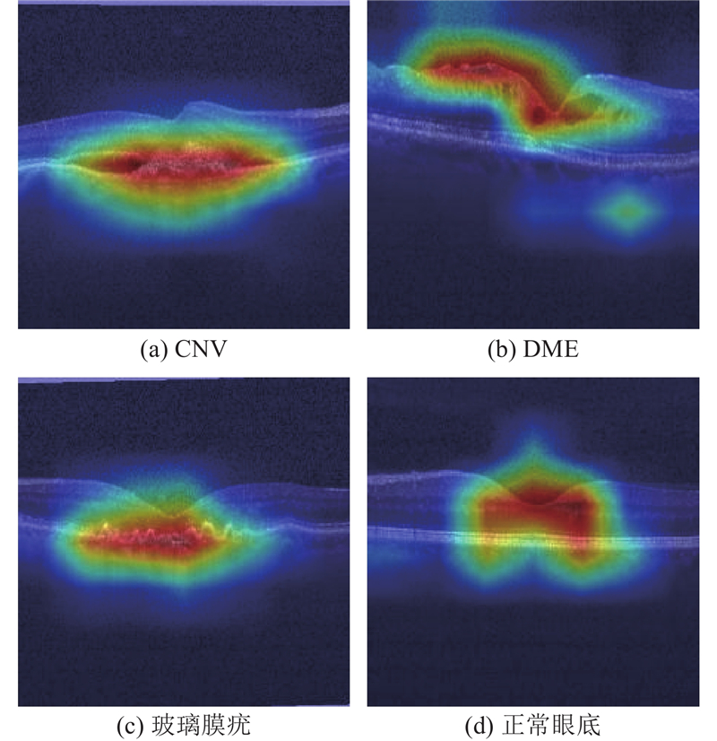
Abstract A lightweight classification model MB-CNN for optical coherence tomography (OCT) images was proposed to accurately and conveniently identify multiple types of fundus lesions. By reducing the number of convolution cores and adjusting the proportion of convolution blocks in each stage, a lightweight backbone network L-Resnet was designed, and the extraction of deep-layer semantic information was enhanced by deepening the network depth. The multi-scale convolution block MultiBlock was designed using depthwise seperable convolution, and the features of the lesion area was mined. Different convolution kernels were used to extract the lesions features of different sizes to improve the recognition ability of the network to the OCT image of the lesion. The feature fusion module FFM was constructed, and the shallow layer information and deep layer information were fused, the texture and semantic information of the pathological features were extracted, and the recognition ability of small target lesions was improved. Experimental result showed that the overall classification accuracy of MB-CNN in the three datasets of UCSD, Duke and NEH was 97.2%, 99.92% and 94.37% respectively, the amount of model parameters were significantly reduced. The proposed model can classify various fundus lesions.
|
|
Received: 23 March 2023
Published: 27 December 2023
|
|
|
| Fund: 安徽理工大学医学专项培育项目(YZ2023H2B006);安徽理工大学引进人才科研启动基金资助项目(2022yjrc44);国家自然科学基金资助项目(52174141);安徽省自然科学基金资助项目(2108085ME158);安徽理工大学研究生创新基金资助项目(2022CX2086) |
|
Corresponding Authors:
Xiao-fen JIA
E-mail: hxh19855424153@163.com;jxfzbt2008@163.com
|
眼底病变OCT图像的轻量化识别算法
为了准确、方便地识别多类型眼底病变,提出光学相干断层扫描技术(OCT)图像的轻量化分类模型MB-CNN. 降低卷积核的使用个数,调节每个阶段卷积块的使用比例,设计轻量化主干网络L-Resnet,通过加深网络深度增强对深层语义信息的提取. 使用深度可分离卷积设计多尺度卷积块MultiBlock,利用MultiBloc深度挖掘病灶区域的特征,使用不同的卷积核提取不同尺寸病变的特征,提高网络对病变OCT图像的识别能力. 构建特征融合模块FFM,融合浅层信息和深层信息,充分提取病变特征的纹理和语义信息,提高对小目标病变的识别能力. 实验结果显示,MB-CNN在UCSD、Duke和NEH3个数据集上的总体分类精度分别达到97.2%、99.92%和94.37%,模型参数量明显降低,所提模型能够针对眼底的多种病变进行分类.
关键词:
眼底病变,
光学相干断层扫描技术(OCT)图像,
智能识别,
轻量化分类模型,
语义信息,
特征融合
|
|
| [1] |
FERRIS III F L, WILKINSON C P, BIRD A, et al Clinical classification of age-related macular degeneration[J]. Ophthalmology, 2013, 120 (4): 844- 851
doi: 10.1016/j.ophtha.2012.10.036
|
|
|
| [2] |
张昊瑞, 桂潇, 赵娜, 等 糖尿病性黄斑水肿的药物治疗研究进展[J]. 中国眼耳鼻喉科杂志, 2021, 21 (3): 226- 229
ZHANG Hao-rui, GUI Xiao, ZHAO Na, et al Research progress on pharmacotherapy for diabetic macular edema[J]. Chinese Journal of Ophthalmology and Otorhinolaryngology, 2021, 21 (3): 226- 229
|
|
|
| [3] |
HUANG D, SWANSON E A, LIN C P, et al Optical coherence tomography[J]. Science, 1991, 254 (5035): 1178- 1181
doi: 10.1126/science.1957169
|
|
|
| [4] |
PODOLEANU A G Optical coherence tomography[J]. Journal of Microscopy, 2012, 247 (3): 209- 219
doi: 10.1111/j.1365-2818.2012.03619.x
|
|
|
| [5] |
FANG L, WANG C, LI S, et al Attention to lesion: lesion-aware convolutional neural network for retinal optical coherence tomography image classification[J]. IEEE Transactions on Medical Imaging, 2019, 38 (8): 1959- 1970
doi: 10.1109/TMI.2019.2898414
|
|
|
| [6] |
LIU X, BAI Y, CAO J, et al Joint disease classification and lesion segmentation via one-stage attention-based convolutional neural network in OCT images[J]. Biomedical Signal Processing and Control, 2022, 71: 103087
doi: 10.1016/j.bspc.2021.103087
|
|
|
| [7] |
MISHRA S S, MANDAL B, PUHAN N B Perturbed composite attention model for macular optical coherence tomography image classification[J]. IEEE Transactions on Artificial Intelligence, 2022, 3 (4): 625- 635
doi: 10.1109/TAI.2021.3135797
|
|
|
| [8] |
DAS V, DANDAPAT S, BORA P K Automated classification of retinal OCT images using a deep multi-scale fusion CNN[J]. IEEE Sensors Journal, 2021, 21 (20): 23256- 23265
doi: 10.1109/JSEN.2021.3108642
|
|
|
| [9] |
THOMAS A, HARIKRISHNAN P M, KRISHAN A K, et al A novel multiscale convolutional neural network based age-related macular degeneration detection using OCT images[J]. Biomedical Signal Processing and Control, 2021, 67: 102538
doi: 10.1016/j.bspc.2021.102538
|
|
|
| [10] |
TOĞAÇAR M, ERGEN B, TÜMEN V Use of dominant activations obtained by processing OCT images with the CNNs and slime mold method in retinal disease detection[J]. Biocybernetics and Biomedical Engineering, 2022, 42 (2): 646- 666
doi: 10.1016/j.bbe.2022.05.005
|
|
|
| [11] |
SOTOUDEH-PAIMA S, JODEIRI A, HAJIZADEH F, et al Multi-scale convolutional neural network for automated AMD classification using retinal OCT images[J]. Computers in Biology and Medicine, 2022, 144: 105368
doi: 10.1016/j.compbiomed.2022.105368
|
|
|
| [12] |
HE K, ZHANG X, REN S, et al. Deep residual learning for image recognition [C]// Proceedings of the 2016 IEEE Conference on Computer Vision and Pattern Recognition. Las Vegas: IEEE, 2016: 770-778.
|
|
|
| [13] |
HE K, ZHANG X, REN S, et al. Identity mappings in deep residual networks [C]// Computer Vision–ECCV 2016. [S.l.]: Springer, 2016: 630-645.
|
|
|
| [14] |
LIU Z, MAO H, WU C, et al. A convnet for the 2020s [C]// Proceedings of the 2022 IEEE/CVF Conference on Computer Vision and Pattern Recognition. New Orleans: IEEE, 2022: 11976-11986.
|
|
|
| [15] |
SZEGEDY C, LIU W, JIA Y, et al. Going deeper with convolutions [C]// Proceedings of the 2015 IEEE Conference on Computer Vision and Pattern Recognition. Boston: IEEE, 2015: 1-9.
|
|
|
| [16] |
CHOLLET F. Xception: deep learning with depthwise separable convolutions [C]// Proceedings of the 2017 IEEE Conference on Computer Vision and Pattern Recognition. Honolulu: IEEE, 2017: 1251-1258.
|
|
|
| [17] |
HAN K, WANG Y, TIAN Q, et al. GhostNet: more features from cheap operations [C]// Proceedings of the 2020 IEEE/CVF Conference on Computer Vision and Pattern Recognition. Seattle: IEEE, 2020: 1580-1589.
|
|
|
| [18] |
IOFFE S, SZEGEDY C. Batch normalization: accelerating deep network training by reducing internal covariate shift[C]// Proceeding of the 32nd International Conference on Machine Learning.[S.l.]: PMLR, 2015: 448-456.
|
|
|
| [19] |
KERMANY D S, GOLDBAUM M, CAI W, et al Identifying medical diagnoses and treatable diseases by image-based deep learning[J]. Cell, 2018, 172 (5): 1122- 1131
doi: 10.1016/j.cell.2018.02.010
|
|
|
|
Viewed |
|
|
|
Full text
|
|
|
|
|
Abstract
|
|
|
|
|
Cited |
|
|
|
|
| |
Shared |
|
|
|
|
| |
Discussed |
|
|
|
|