| 计算机技术 |
|

|
|
|
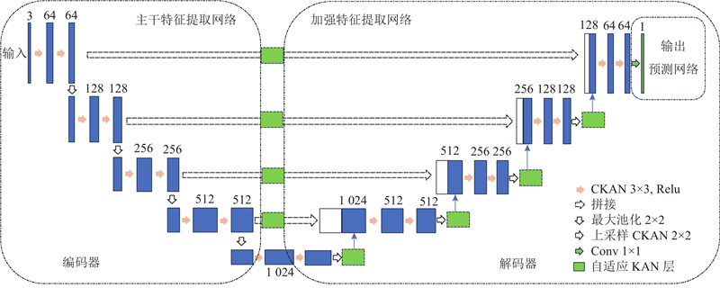
| 基于KAN与CKAN优化的医学图像分割模型 |
娄世猛1( ),邵玉斌1,*(),杜庆治1,唐菁敏1,张赜涛2 ),邵玉斌1,*(),杜庆治1,唐菁敏1,张赜涛2 |
1. 昆明理工大学 信息工程与自动化学院,云南 昆明 650500
2. 云南省媒体融合重点实验室,云南 昆明 650228 |
|
| Medical image segmentation model based on KAN and CKAN optimization |
| Shimeng LOU1(),Yubin SHAO1,*(),Qingzhi DU1,Jingmin TANG1,Zetao ZHANG2 |
1. Faculty of Information Engineering and Automation, Kunming University of Science and Technology, Kunming 650500, China
2. Yunnan Province Key Laboratory for Media Integration, Kunming 650228, China |
引用本文:
娄世猛,邵玉斌,杜庆治,唐菁敏,张赜涛. 基于KAN与CKAN优化的医学图像分割模型[J]. 浙江大学学报(工学版), 2026, 60(6): 1277-1288.
Shimeng LOU,Yubin SHAO,Qingzhi DU,Jingmin TANG,Zetao ZHANG. Medical image segmentation model based on KAN and CKAN optimization. Journal of ZheJiang University (Engineering Science), 2026, 60(6): 1277-1288.
链接本文:
https://www.zjujournals.com/eng/CN/10.3785/j.issn.1008-973X.2026.06.015
或
https://www.zjujournals.com/eng/CN/Y2026/V60/I6/1277
|
| 1 |
GARCIA-GARCIA A, ORTS-ESCOLANO S, OPREA S, et al. A review on deep learning techniques applied to semantic segmentation [EB/OL]. [2025-08-17]. https://arxiv.org/abs/1704.06857.
|
| 2 |
MINAEE S, BOYKOV Y, PORIKLI F, et al Image segmentation using deep learning: a survey[J]. IEEE Transactions on Pattern Analysis and Machine Intelligence, 2021, 44 (7): 3523- 3542
|
| 3 |
ROTH H R, LU L, FARAG A, et al. DeepOrgan: multi-level deep convolutional networks for automated pancreas segmentation [C]//Medical Image Computing and Computer-Assisted Intervention. Cham: Springer, 2015: 556–564.
|
| 4 |
LITJENS G, KOOI T, BEJNORDI B E, et al A survey on deep learning in medical image analysis[J]. Medical Image Analysis, 2017, 42: 60- 88
doi: 10.1016/j.media.2017.07.005
|
| 5 |
DEVALLA S K, PHAM T H, PANDA S K, et al Towards label-free 3D segmentation of optical coherence tomography images of the optic nerve head using deep learning[J]. Biomedical Optics Express, 2020, 11 (11): 6356- 6378
doi: 10.1364/BOE.395934
|
| 6 |
CORDTS M, OMRAN M, RAMOS S, et al. The cityscapes dataset for semantic urban scene understanding [C]//Proceedings of the IEEE Conference on Computer Vision and Pattern Recognition. Las Vegas: IEEE, 2016: 3213–3223.
|
| 7 |
RONNEBERGER O, FISCHER P, BROX T. U-Net: convolutional networks for biomedical image segmentation [C]//Medical Image Computing and Computer-Assisted Intervention. Cham: Springer, 2015: 234–241.
|
| 8 |
ZHOU Z, RAHMAN SIDDIQUEE M M, TAJBAKHSH N, et al. UNet++: a nested U-Net architecture for medical image segmentation [C]//Deep Learning in Medical Image Analysis and Multimodal Learning for Clinical Decision Support. Cham: Springer, 2018: 3–11.
|
| 9 |
ZHAO H, SHI J, QI X, et al. Pyramid scene parsing network [C]//Proceedings of the IEEE Conference on Computer Vision and Pattern Recognition. Honolulu: IEEE, 2017: 6230–6239.
|
| 10 |
CHEN L C, PAPANDREOU G, KOKKINOS I, et al DeepLab: semantic image segmentation with deep convolutional nets, atrous convolution, and fully connected CRFs[J]. IEEE Transactions on Pattern Analysis and Machine Intelligence, 2018, 40 (4): 834- 848
doi: 10.1109/TPAMI.2017.2699184
|
| 11 |
CAO H, WANG Y, CHEN J, et al. Swin-Unet: Unet-like pure transformer for medical image segmentation [C]// European Conference on Computer Vision. Cham: Springer, 2023: 205–218.
|
| 12 |
ISENSEE F, JAEGER P F, KOHL S A A, et al nnU-NET: a self-configuring method for deep learning-based biomedical image segmentation[J]. Nature Methods, 2021, 18 (2): 203- 211
doi: 10.1038/s41592-020-01008-z
|
| 13 |
ISENSEE F, PETERSEN J, KLEIN A, et al. nnU-NET: self-adapting framework for U-Net-based medical image segmentation [EB/OL]. [2025-08-17]. https://arxiv.org/abs/1809.10486.
|
| 14 |
MOU L, ZHAO Y, CHEN L, et al. CS-Net: channel and spatial attention network for curvilinear structure segmentation [C]//Medical Image Computing and Computer Assisted Intervention. Cham: Springer, 2019: 721–730.
|
| 15 |
STAAL J, ABRAMOFF M D, NIEMEIJER M, et al Ridge-based vessel segmentation in color images of the retina[J]. IEEE Transactions on Medical Imaging, 2004, 23 (4): 501- 509
doi: 10.1109/TMI.2004.825627
|
| 16 |
LIU Z, WANG Y, VAIDYA S, et al. KAN: Kolmogorov-Arnold networks [EB/OL]. [2025-08-17]. https://arxiv.org/abs/2404.19756.
|
| 17 |
BODNER A D, TEPSICH A S, SPOLSKI J N, et al. Convolutional Kolmogorov-Arnold networks [EB/OL]. [2025-08-17]. https://arxiv.org/abs/2406.13155.
|
| 18 |
LI C, LIU X, LI W, et al U-KAN makes strong backbone for medical image segmentation and generation[J]. Proceedings of the AAAI Conference on Artificial Intelligence, 2025, 39 (5): 4652- 4660
doi: 10.1609/aaai.v39i5.32491
|
| 19 |
MA X, WANG Z, HU Y, et al. Kolmogorov-Arnold network for remote sensing image semantic segmentation [EB/OL]. [2025-08-17]. https://arxiv.org/abs/2501.07390.
|
| 20 |
AGRAWAL A, AGRAWAL A, GUPTA S, et al. KAN-Mamba FusionNet: redefining medical image segmentation with non-linear modeling [EB/OL]. [2025-08-17]. https://arxiv.org/abs/2411.11926.
|
| 21 |
OKTAY O, SCHLEMPER J, FOLGOC L L, et al. Attention U-Net: learning where to look for the pancreas [EB/OL]. [2025-08-17]. https://arxiv.org/abs/1804.03999.
|
| 22 |
ZHANG Z, LIU Q, WANG Y Road extraction by deep residual U-Net[J]. IEEE Geoscience and Remote Sensing Letters, 2018, 15 (5): 749- 753
doi: 10.1109/LGRS.2018.2802944
|
| 23 |
BILIC P, CHRIST P, LI H B, et al The Liver tumor segmentation benchmark (LiTS)[J]. Medical Image Analysis, 2023, 84: 102680
doi: 10.1016/j.media.2022.102680
|
| 24 |
SCHOENBERG I J. Contributions to the problem of approximation of equidistant data by analytic functions: part A. -on the problem of smoothing or graduation. a first class of analytic approximation formulae [J]. Quarterly of Applied Mathematics, 1946, 4(1): 45–99.
|
| 25 |
HOLLADAY J C A smoothest curve approximation[J]. Mathematical Tables and Other Aids to Computation, 1957, 11 (60): 233- 243
doi: 10.1090/s0025-5718-1957-0093894-6
|
| 26 |
MILLETARI F, NAVAB N, AHMADI S A. V-net: fully convolutional neural networks for volumetric medical image segmentation [C]// Proceedings of the Fourth International Conference on 3D Vision. Stanford: IEEE, 2016: 565–571.
|
| 27 |
EVERINGHAM M, VAN GOOL L, WILLIAMS C K I, et al The pascal visual object classes (VOC) challenge[J]. International Journal of Computer Vision, 2010, 88 (2): 303- 338
doi: 10.1007/s11263-009-0275-4
|
| 28 |
TAGHANAKI S A, ZHENG Y, ZHOU K S, et al Combo loss: handling input and output imbalance in multi-organ segmentation[J]. Computerized Medical Imaging and Graphics, 2019, 75: 24- 33
doi: 10.1016/j.compmedimag.2019.04.005
|
|
Viewed |
|
|
|
Full text
|
|
|
|
|
Abstract
|
|
|
|
|
Cited |
|
|
|
|
| |
Shared |
|
|
|
|
| |
Discussed |
|
|
|
|