| 生物医学工程 |
|


|
|
|
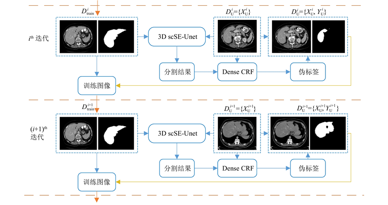
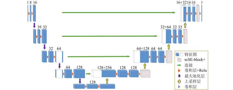
| 基于3D scSE-UNet的肝脏CT图像半监督学习分割方法 |
刘清清1,2( ),周志勇2,范国华3,钱旭升1,2,胡冀苏1,2,陈光强3,戴亚康2,4,*() ),周志勇2,范国华3,钱旭升1,2,胡冀苏1,2,陈光强3,戴亚康2,4,*() |
1. 中国科学技术大学 生命科学与医学部 生物医学工程学院(苏州),江苏 苏州 215163
2. 中国科学院 苏州生物医学工程技术研究所,江苏 苏州 215163
3. 苏州大学附属第二医院,江苏 苏州 215000
4. 济南国科医工科技发展有限公司,山东 济南 250000 |
|
| Semi-supervised learning segmentation method of liver CT images based on 3D scSE-UNet |
| Qing-qing LIU1,2(),Zhi-yong ZHOU2,Guo-hua FAN3,Xu-sheng QIAN1,2,Ji-su HU1,2,Guang-qiang CHEN3,Ya-kang DAI2,4,*() |
1. School of Biomedical Engineering (Suzhou), Division of Life Sciences and Medicine, University of Science and Technology of China, Suzhou 215163, China
2. Suzhou Institute of Biomedical Engineering and Technology, Chinese Academy of Science, Suzhou 215163, China
3. The Second Affiliated Hospital of Suzhou University, Suzhou 215000, China
4. Jinan Guoke Medical Engineering Technology Development Limited Company, Jinan 250000, China |
引用本文:
刘清清,周志勇,范国华,钱旭升,胡冀苏,陈光强,戴亚康. 基于3D scSE-UNet的肝脏CT图像半监督学习分割方法[J]. 浙江大学学报(工学版), 2021, 55(11): 2033-2044.
Qing-qing LIU,Zhi-yong ZHOU,Guo-hua FAN,Xu-sheng QIAN,Ji-su HU,Guang-qiang CHEN,Ya-kang DAI. Semi-supervised learning segmentation method of liver CT images based on 3D scSE-UNet. Journal of ZheJiang University (Engineering Science), 2021, 55(11): 2033-2044.
链接本文:
https://www.zjujournals.com/eng/CN/10.3785/j.issn.1008-973X.2021.11.003
或
https://www.zjujournals.com/eng/CN/Y2021/V55/I11/2033
|
| 1 |
LEO J Computer-aided surgery meets predictive, preventive and personalized medicine[J]. EPMA Journal, 2017, 8 (1): 1- 4
doi: 10.1007/s13167-017-0084-8
|
| 2 |
ZHOU S, CHENG Y, TAMURA S Automated lung seg- mentation and smoothing techniques for inclusion of juxtapleural nodules and pulmonary vessels on chest CT images[J]. Biomedical Signal Processing and Control, 2014, 13: 62- 70
doi: 10.1016/j.bspc.2014.03.010
|
| 3 |
SHI C, CHENG Y, WANG J, et al Low-rank and sparse decomposition based shape model and probabilistic atlas for automatic pathological organ segmentation[J]. Medical Image Analysis, 2017, 38: 30- 49
doi: 10.1016/j.media.2017.02.008
|
| 4 |
SHI C, CHENG Y, LIU F, et al A hierarchical local region-based sparse shape composition for liver segmentation in CT scans[J]. Pattern Recognition, 2016, 50: 88- 106
doi: 10.1016/j.patcog.2015.09.001
|
| 5 |
LI G, CHEN X, SHI F, et al Automatic liver segmentation based on shape constraints and deformable graph cut in CT images[J]. IEEE Transactions on Image Processing A Publication of the IEEE Signal Processing Society, 2015, 24 (12): 5315
doi: 10.1109/TIP.2015.2481326
|
| 6 |
LIU Z, SONG Y, SHENG V, et al Liver CT sequence segmentation based with improved U-Net and graph cut[J]. Expert Systems with Applications, 2019, 126: 54- 63
doi: 10.1016/j.eswa.2019.01.055
|
| 7 |
LU X, WU J, REN X, et al The study and application of the improved region growing algorithm for liver segmentation[J]. Optik-International Journal for Light and Electron Optics, 2014, 125 (9): 2142- 2147
doi: 10.1016/j.ijleo.2013.10.049
|
| 8 |
RAFIEI S, KARIMI N, MIRMAHBOUB B, et al. Liver segmentation in abdominal CT images using probabilistic atlas and adaptive 3D region growing [C]// 2019 41st Annual International Conference of the IEEE Engineering in Medicine and Biology Society (EMBC). Berlin: IEEE, 2019.
|
| 9 |
郑洲, 张学昌, 郑四鸣, 等 基于区域增长与统一化水平集的CT肝脏图像分割[J]. 浙江大学学报: 工学版, 2018, 52 (12): 145- 159
ZHENG Zhou, ZHANG Xue-chang, ZHENG Si-ming, et al Liver segmentation in CT images based on region-growing and unified level set method[J]. Journal of Zhejiang University: Engineering Science, 2018, 52 (12): 145- 159
|
| 10 |
WANG J, CHENG Y, GUO C, et al Shape-intensity prior level set: combining probabilistic atlas and probability map constrains for automatic liver segmentation from abdominal CT images[J]. International Journal of Computer Assisted Radiology and Surgery, 2016, 11 (5): 817- 826
doi: 10.1007/s11548-015-1332-9
|
| 11 |
LI X, CHEN H, QI X, et al H-DenseUNet: hybrid densely connected UNet for liver and tumor segmentation from CT volumes[J]. IEEE Transactions on Medical Imaging, 2018, 37 (12): 2663- 2674
doi: 10.1109/TMI.2018.2845918
|
| 12 |
LONG J, SHELHAMER E, DARRELL T Fully convolutional networks for semantic segmentation[J]. IEEE Transactions on Pattern Analysis and Machine Intelligence, 2015, 39 (4): 640- 651
|
| 13 |
孙明建, 徐军, 马伟, 等 基于新型深度全卷积网络的肝脏CT影像三维区域自动分割[J]. 中国生物医学工程学报, 2018, 37 (4): 385- 393
SUN Ming-jian, XU Jun, MA Wei, et al A new fully convolutional network for 3D liver region segmentation on CT images[J]. Chinese Journal of Biomedical Engineering, 2018, 37 (4): 385- 393
doi: 10.3969/j.issn.0258-8021.2018.04.001
|
| 14 |
RONNEBERGER O, FISCHER P, BROX T. U-Net: convolutional networks for biomedical image segmentation [C]// International Conference on Medical Image Computing and Computer Assisted Intervention. Munich: Springer, 2015: 234-241.
|
| 15 |
ÇIÇEK Ö, ABDULKADIR A, LIENKAMP S, et al. 3D U-Net: learning dense volumetric segmentation from sparse annotation [C]// International Conference on Medical Image Computing and Computer-Assisted Intervention. Athens: Springer, 2016: 424-432.
|
| 16 |
CHRIST P, ELSHAER M, ETTLINGER F, et al. Automatic liver and lesion segmentation in CT using cascaded fully convolutional neural networks and 3D conditional random fields [C]// International Conference on Medical Image Computing and Computer-Assisted Intervention. Athens: Springer, 2016: 415-423.
|
| 17 |
刘哲, 张晓林, 宋余庆, 等 结合改进的U-Net和Morphsnakes的肝脏分割[J]. 中国图象图形学报, 2018, 23 (8): 1254- 1262
LIU Zhe, ZHANG Xiao-lin, SONG Yu-qing, et al Liver segmentation with improved U-Net and Morphsnakes algorithm[J]. Journal of Image and Graphics, 2018, 23 (8): 1254- 1262
|
| 18 |
HE F, ZHANG G, YANG H, et al Multi-scale attention module U-Net liver tumour segmentation method[J]. Journal of Physics: Conference Series, 2020, 1678: 012107
doi: 10.1088/1742-6596/1678/1/012107
|
| 19 |
OKTAY O, SCHLEMPER J, FOLGOC L, et al. Attention U-Net: learning where to look for the pancreas [EB/OL]. [2020-11-20]. https://arxiv.org/pdf/1804.03999.
|
| 20 |
HU J, SHEN L, SUN G, et al. Squeeze-and-excitation networks [C]// Proceedings of the IEEE conference on Computer Vision and Pattern Recognition. Salt Lake City: IEEE, 2018: 7132-7141.
|
| 21 |
ROY A, NAVAB N, WACHINGER C. Concurrent spatial and channel ‘squeeze & excitation’ in fully convolutional networks [C]// International Conference on Medical Image Computing and Computer Assisted Intervention. Granada: Springer, 2018, 421-429.
|
| 22 |
潘崇煜, 黄健, 郝建国, 等 融合零样本学习和小样本学习的弱监督学习方法综述[J]. 系统工程与电子技术, 2020, 42 (10): 2246- 2256
PAN Chong-yu, HUANG Jian, HAO Jian-guo, et al Survey of weakly supervised learning integrating zero-shot and few-shot learing[J]. Systems Engineering and Electionics, 2020, 42 (10): 2246- 2256
doi: 10.3969/j.issn.1001-506X.2020.10.13
|
| 23 |
NAKAYAMA Y, LI Q, KATSURAGAWA S, et al Automated hepatic volumetry for living related liver transplantation at multisection CT[J]. Radiology, 2006, 240 (3): 743- 748
doi: 10.1148/radiol.2403050850
|
| 24 |
CHEPLYGINA V, DE BRUIJNE M, PLUIM J Not-so- supervised: a survey of semi-supervised, multi-instance, and transfer learning in medical image analysis[J]. Medical Image Analysis, 2019, 54: 280- 296
doi: 10.1016/j.media.2019.03.009
|
| 25 |
XIE Q, LUONG M, HOVY E, et al. Self-training with noisy student improves imagenet classification [C]// Proceedings of the IEEE/CVF Conference on Computer Vision and Pattern Recognition. [s.l.]: IEEE, 2020, 10687-10698.
|
| 26 |
ZHAO F, CHEN Y, CHEN F, et al Semi-supervised cerebrovascular segmentation by hierarchical convolutional neural network[J]. IEEE Access, 2018, 6: 67841- 67852
doi: 10.1109/ACCESS.2018.2879521
|
| 27 |
XIA, Y, LIU F, YANG D, et al. 3D Semi-supervised learning with uncertainty-aware multi-view co-training [C]// 2020 Workshop on Applications of Computer Vision. Snowmass Village: IEEE, 2020: 3646-3655.
|
| 28 |
YANG Z, COHEN W, SALAKHUTDINOV R. Revisiting semi-supervised learning with graph embeddings [C]// International Conference on Machine Learning. New York City: JMLR, 2016: 40-48.
|
| 29 |
ZHOU Y, WANG Y, TANG P, et al. Semi-supervised 3D abdominal multi-organ segmentation via deep multi-planar co-training [C]// 2019 IEEE Winter Conference on Applications of Computer Vision (WACV). Hawaii: IEEE, 2019: 121-140.
|
| 30 |
JIANG B, ZHANG Z, LIN D, et al. Semi-supervised learning with graph learning-convolutional networks [C]// Proceedings of the IEEE conference on Computer Vision and Pattern Recognition. Long Beach: IEEE,, 2019: 11313-11320.
|
| 31 |
姜威威, 刘祥强, 韩金仓 基于深度协同训练的肝脏CT图像自动分割方法[J]. 电子设计工程, 2020, 28 (14): 175- 179
JIANG Wei-wei, LIU Xiang-qiang, HAN Jin-cang Automatic liver segmentation from CT images based on deep co-training[J]. Electronic Design Engineering, 2020, 28 (14): 175- 179
|
| 32 |
LEE D. Pseudo-label: the simple and efficient semi-supervised learning method for deep neural networks [C]// In Workshop on Challenges in Representation Learning, ICML. Atlanta: JMLR, 2013, 3: 896.
|
| 33 |
BAI W, OKTAY O, SINCLAIR M, et al. Semi-supervised learning for network-based cardiac MR image segmentation [C]// International Conference on Medical Image Computing and Computer Assisted Intervention. Quebec: Springer, 2017: 253-260.
|
| 34 |
KRAHENBUHL P, KOLTUN V Efficient inference in fully connected CRFs with Gaussian edge potentials[J]. Advances in Neural Information Processing Systems, 2011, 24: 109- 117
|
|
Viewed |
|
|
|
Full text
|
|
|
|
|
Abstract
|
|
|
|
|
Cited |
|
|
|
|
| |
Shared |
|
|
|
|
| |
Discussed |
|
|
|
|