|
|
|
| Classification method for electrocardiograph signals based on parallel architecture model and spatial-temporal attention mechanism |
Xiang-dong PENG( ),Cong-cheng PAN(),Ze-jun KE,Hua-qiang ZHU,Xiao ZHOU ),Cong-cheng PAN(),Ze-jun KE,Hua-qiang ZHU,Xiao ZHOU |
| School of Software and Internet of Things Engineering, Jiangxi University of Finance and Economics, Nanchang 330032, China |
|
|
|
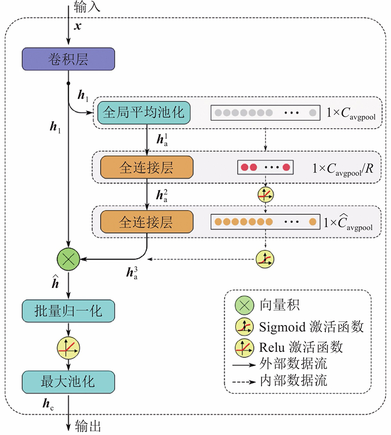
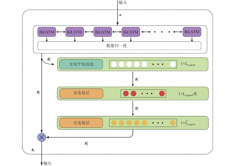
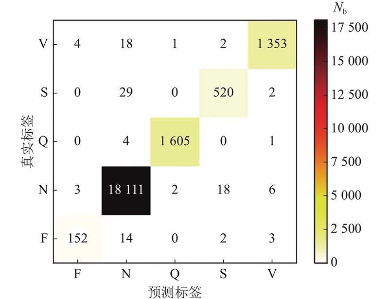
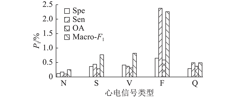
Abstract A parallel architecture electrocardiograph (ECG) classification model based on deep learning was proposed in order to effectively extract the spatiotemporal characteristics of ECG signals and improve the classification accuracy. A spatiotemporal attention mechanism based on gate channel attention block (GCA block) and gate time step attention (GTSA block) module was adopted in order to achieve multi-channel feature fusion. The bidirectional long-short time memory network and the convolutional neural network were used as the base feature extractor. The before-after dependence of the ECG signal sequence data and the local correlation features at different scales were captured respectively, and the automatic classification of five different types of ECG signals was realized. Results verified on the MIT-BIH dataset showed that the accuracy, specificity, sensitivity, accuracy and Macro-F1 of the total classification of five different ECG signals by the method were 99.50%, 99.61%, 96.20%, 98.02% and 97.08%, respectively. The model can not only effectively shorten the depth of the network model and prevent the model from overfitting, but also more accurately extract the spatiotemporal characteristics of the ECG signal and obtain better classification performance compared with other ECG classification models.
|
|
Received: 06 March 2022
Published: 25 October 2022
|
|
|
| Fund: 江西省自然科学基金资助项目(20192BAB207003);江西省教育厅科学技术研究资助项目(GJJ180263) |
基于并行架构和时空注意力机制的心电分类方法
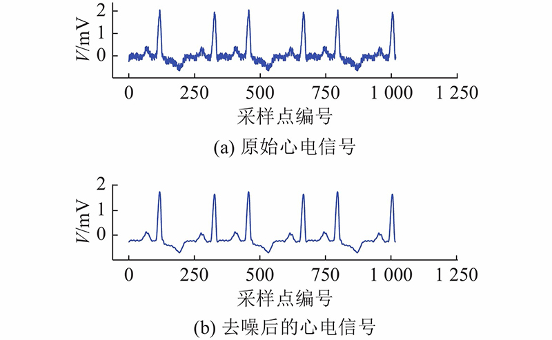
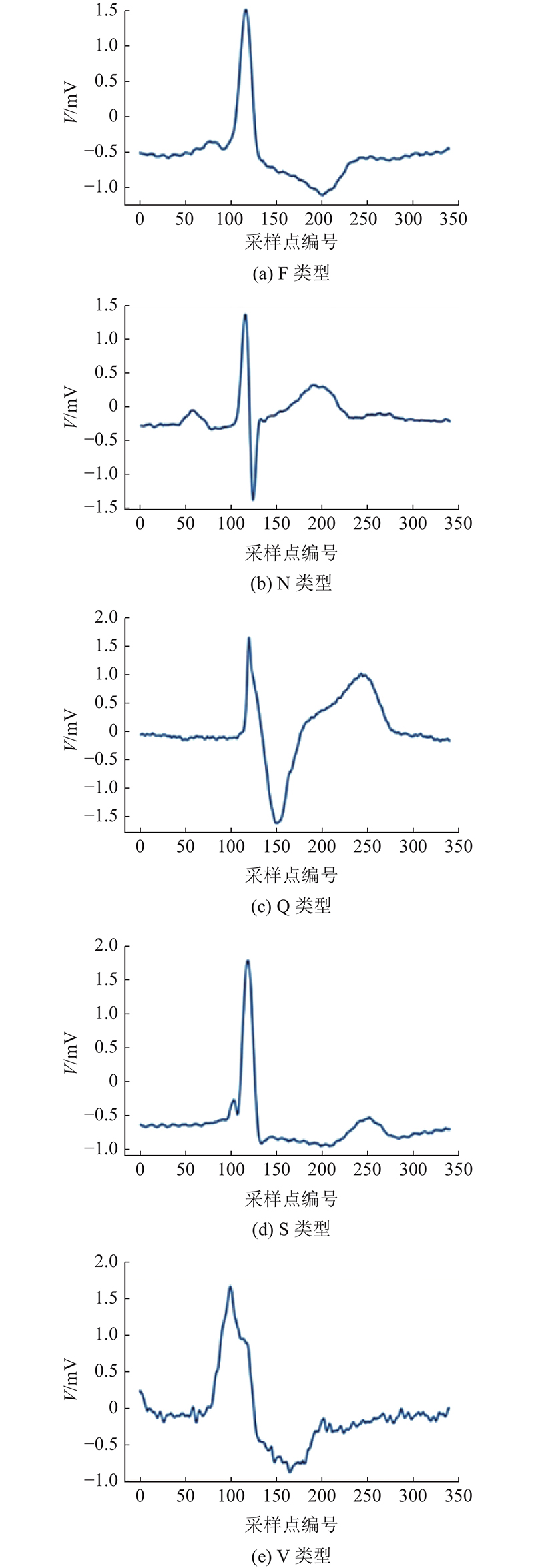
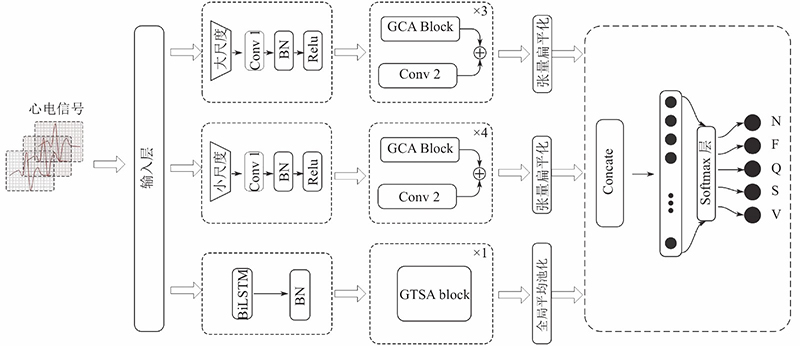
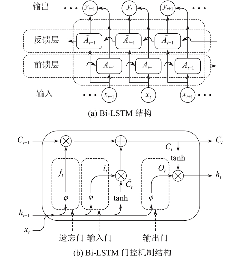
为了有效提取心电信号 (ECG) 的时空特征和提高分类准确性,提出基于深度学习的并行架构心电分类模型. 该模型采用基于GCA Block和GTSA Block模块实现多路特征融合的时空注意力机制. 使用双向长短时记忆网络和卷积神经网络作为基特征提取器,分别捕捉心电信号序列数据的前后依赖关系和不同尺度上的局部相关特征,实现对5种不同类型的心电信号的自动分类. 在MIT-BIH数据集上验证的结果表明,该方法对5种不同心电信号的总体分类准确率、特异性、敏感度、精确度和Macro-F1分别为99.50%、99.61%、96.20%、98.02%和97.08%. 相较于其他心电分类模型,该模型不仅能够有效地缩短网络模型深度,防止模型过拟合,而且能够更准确地提取心电信号的时空特征,获得更好的分类性能.
关键词:
心电分类,
数据不平衡,
深度学习,
并行架构,
时空注意力机制
|
|
| [1] |
EBRAHIMI Z, LONI M, DANESHTALAB M, et al A review on deep learning methods for ECG arrhythmia classification[J]. Expert Systems with Applications: X, 2020, 7: 100033
doi: 10.1016/j.eswax.2020.100033
|
|
|
| [2] |
CHOI S, ADNANE M, LEE G J, et al Development of ECG beat segmentation method by combining low-pass filter and irregular R–R interval checkup strategy[J]. Expert Systems with Applications, 2010, 37 (7): 5208- 5218
doi: 10.1016/j.eswa.2009.12.069
|
|
|
| [3] |
ASL B, SETAREHADN S, MOHEBBI M Support Vector machine-based arrhythmia classification using reduced features of heart rate variability signal[J]. Artificial Intelligence in Medicine, 2008, 44 (1): 51- 64
doi: 10.1016/j.artmed.2008.04.007
|
|
|
| [4] |
FAN X, YAO Q, CAI Y, et al Multi-scaled fusion of deep convolutional neural networks for screening atrial fibrillation from single lead short ecg recordings[J]. IEEE Journal of Biomedical and Health Informatics, 2018, 22 (6): 1744- 1753
doi: 10.1109/JBHI.2018.2858789
|
|
|
| [5] |
HANNUN A Y, RAJPURKAR P, HAGHPANAHI M, et al Cardiologist-level arrhythmia detection and classification in ambulatory electrocardiograms using a deep neural network[J]. Nature Medicine, 2019, 25 (1): 65- 69
doi: 10.1038/s41591-018-0268-3
|
|
|
| [6] |
ZHANG J, LIU A, GAO M, et al ECG-based multi-class arrhythmia detection using spatio-temporal attention based convolutional recurrent neural network[J]. Artificial Intelligence in Medicine, 2020, 106: 101856
doi: 10.1016/j.artmed.2020.101856
|
|
|
| [7] |
SAADATNEJAD S, OVEISI M, HASHEMI M LST-M based ECG classification for continuous monitoring on personal wearable devices[J]. IEEE Journal of Biomedical and Health Informatics, 2019, 24 (2): 515- 523
|
|
|
| [8] |
LYNN H M, PAN S B, KIM P A deep bidirectional GRU network model for biometric electrocardiogram classification based on recurrent neural networks[J]. IEEE Access, 2019, 7: 145395- 145405
|
|
|
| [9] |
YAO Q, FAN X, CAI Y, et al. Time-incremental convolutional neural network for arrhythmia detection in varied-length electrocardiogram [C]// 2018 IEEE 16th International Conference on Dependable, Autonomic and Secure Computing, 16th International Conference on Pervasive Intelligence and Computing, 4th International Conference on Big Data Intelligence and Computing and Cyber Science and Technology Congress. Athens: IEEE, 2018: 754-761.
|
|
|
| [10] |
HE R, LIU Y, WANG K, et al Automatic cardiac arrhythmia classification using combination of deep residual network and bidirectional LSTM[J]. IEEE Access, 2019, 7: 102119- 102135
doi: 10.1109/ACCESS.2019.2931500
|
|
|
| [11] |
YAO Q, WANG R, FAN X, et al Multi-class arrhythmia detection from 12-lead varied-length ECG using attention based time-incremental convolutional neural network[J]. Information Fusion, 2020, 53: 174- 182
doi: 10.1016/j.inffus.2019.06.024
|
|
|
| [12] |
WANG P, HOU B, SHAO S, et al ECG arrhythmiasdetection using auxiliary classifier generative adversarial network and residual network[J]. IEEE Access, 2019, 27 (2): 100910- 100922
|
|
|
| [13] |
HOU B, YANG J, WANG P, et al LSTM-based auto-encoder model for ECG arrhythmias classification[J]. IEEE Transactions on Instrumentation and Measurement, 2019, 69 (4): 1232- 1240
|
|
|
| [14] |
MOODY G B, MARK R G The impact of the MIT-BIH arrhythmia database[J]. IEEE Engineering in Medicine and Biology Magazine, 2001, 20 (3): 45- 50
doi: 10.1109/51.932724
|
|
|
| [15] |
ZHANG M, LU C, LIU C Improved double-threshold denoising method based on the wavelet transform[J]. OSA Continuum, 2019, 2 (8): 2328- 2342
doi: 10.1364/OSAC.2.002328
|
|
|
| [16] |
ANSI/AAMI. Testing and reporting performance results of cardiac rhythm and ST segment measurement algorithms[S]. Washington: ANSI, 2008.
|
|
|
| [17] |
HE H, BAI Y, GARCIA E A, et al. ADASYN: adaptive synthetic sampling approach for imbalanced learning [C]// 2008 IEEE International Joint Conference on Neural Networks. Hongkong: IEEE, 2008: 1322-1328.
|
|
|
| [18] |
XU X, JEONG S, LI J Interpretation of electrocardiogram (ECG) rhythm by combined CNN and BiLSTM[J]. IEEE Access, 2020, 28 (3): 125380- 125388
|
|
|
| [19] |
OH S L, NG E Y K, SAN T R, et al Automated diagnosis of arrhythmia using combination of CNN and LSTM techniques with variable length heart beats[J]. Computers in Biology and Medicine, 2018, 102: 278- 287
doi: 10.1016/j.compbiomed.2018.06.002
|
|
|
| [20] |
HUANG J, CHEN B, YAO B, et al ECG arrhythmia classification using STFT-based spectrogram and convolutional neural network[J]. IEEE Access, 2019, 7: 92871- 92880
doi: 10.1109/ACCESS.2019.2928017
|
|
|
| [21] |
CHEN A, WANG F, LIU W, et al Multi-information fusion neural networks for arrhythmia automatic detection[J]. Computer Methods and Programs in Biomedicine, 2020, 193: 105479
doi: 10.1016/j.cmpb.2020.105479
|
|
|
| [22] |
RAMARAJ E A novel deep learning based gated recurrent unit with extreme learning machine for electrocardiogram (ECG) signal recognition[J]. Biomedical Signal Processing and Control, 2021, 68: 102779
doi: 10.1016/j.bspc.2021.102779
|
|
|
| [23] |
JIN Y, QIN C, HUANG Y, et al Multi-domain modeling of atrial fibrillation detection with twin attentional convolutional long short-term memory neural networks[J]. Knowledge-Based Systems, 2020, 193: 105460
doi: 10.1016/j.knosys.2019.105460
|
|
|
| [24] |
GE R, SHEN T, ZHOU Y, et al Convolutional squeeze-and-excitation network for ECG arrhythmia detection[J]. Artificial Intelligence in Medicine, 2021, 121: 102181
doi: 10.1016/j.artmed.2021.102181
|
|
|
| [25] |
LU Y, JIANG M, WEI L, et al Automated arrhythmia classification using depth wise separable convolutional neural network with focal loss[J]. Biomedical Signal Processing and Control, 2021, 69: 102843
doi: 10.1016/j.bspc.2021.102843
|
|
|
| [26] |
ALJOHANI N R, FAYOUMI A, HASSAN S U A novel focal-loss and class-weight-aware convolutional neural network for the classification of in-text citations[J]. Journal of Information Science, 2021, 6 (3): 165- 176
|
|
|
| [27] |
LUO X, YANG L, CAI H, et al Multi-classification of arrhythmias using a HCR-Net on imbalanced ECG datasets[J]. Computer Methods and Programs in Biomedicine, 2021, 208: 106258
doi: 10.1016/j.cmpb.2021.106258
|
|
|
| [28] |
LI C, ZHAO H, LU W, et al DeepECG: image-based electrocardiogram interpretation with deep convolutional neural networks[J]. Biomedical Signal Processing and Control, 2021, 69: 102824
doi: 10.1016/j.bspc.2021.102824
|
|
|
| [29] |
MURAT F, YILDIRIM O, TALO M, et al Application of deep learning techniques for heartbeats detection using ECG signals analysis and review [J]. Computers in Biology and Medicine, 2020, 120: 103726
doi: 10.1016/j.compbiomed.2020.103726
|
|
|
| [30] |
吴志勇, 丁香乾, 许晓伟, 等 基于深度学习和模糊C均值的心电信号分类方法[J]. 自动化学报, 2018, 44 (1): 1913- 1920
WU Zhi-yong, DING Xiang-qian, XU Xiao-wei, et al A classification method for ECG signals based on deep learning and fuzzy C-means[J]. Journal of Automatica Sinica, 2018, 44 (1): 1913- 1920
doi: 10.16383/j.aas.2018.c170417
|
|
|
| [31] |
GAO J, ZHANG H, LU P, et al An effective LSTM recurrent network to detect arrhythmia on imbalanced ECG dataset[J]. Journal of Healthcare Engineering, 2019, 12 (3): 1- 10
|
|
|
|
Viewed |
|
|
|
Full text
|
|
|
|
|
Abstract
|
|
|
|
|
Cited |
|
|
|
|
| |
Shared |
|
|
|
|
| |
Discussed |
|
|
|
|