|
|
|
| Dual-encoder polyp segmentation with feature-gated fusion and wavelet enhancement |
Liming LIANG( ),Ting KANG,Kangquan CHEN,Yi ZHONG ),Ting KANG,Kangquan CHEN,Yi ZHONG |
| School of Electrical Engineering and Automation, Jiangxi University of Science and Technology, Ganzhou 341000, China |
|
|
|
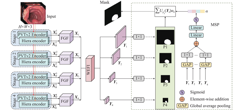
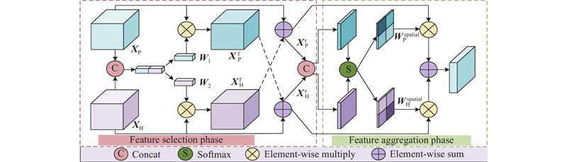
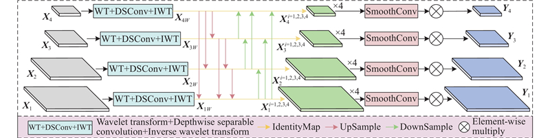
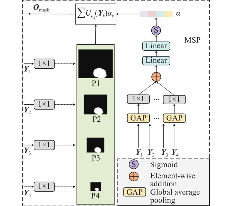
Abstract To address the problems of inaccurate lesion localization, missing boundary details, and small polyp omission in colorectal polyp segmentation, a dual-encoder network with feature-gated fusion and wavelet-enhanced modules was proposed. A dual-encoder architecture based on PVTv2 and Hiera was adopted to enhance global semantic awareness and edge representation. A feature-gated fusion module was designed to filter and combine the multi-level features extracted by two encoders adaptively, thereby better distinguishing polyp regions. A wavelet-enhanced injection module was introduced to decompose high-frequency and low-frequency information using wavelet transform, thus strengthening the edge texture expression and reduced noise, supporting fine-grained feature learning. A multi-scale prediction module was introduced by combining global average pooling and adaptive weighted fusion to achieve refined segmentation and improve adaptability to polyps with diverse shapes. Results show that the proposed network provides effective support for computer-aided diagnosis of colorectal polyps.
|
|
Received: 26 June 2025
Published: 16 July 2026
|
|
|
| Fund: 国家自然科学基金资助项目(51365017,61463018);江西省自然科学基金资助项目(20192BAB205084);江西省教育厅科学技术研究重点项目(GJJ170491,GJJ2200848). |
基于特征门控融合与小波增强的双编码器息肉分割
针对结直肠息肉分割任务中病灶区域定位不准、边界细节缺失及小目标息肉易漏检等问题,提出基于特征门控融合与小波增强的双编码器息肉分割网络. 构建PVTv2与Hiera双编码器,提升全局语义感知能力及增强边缘表征能力. 设计特征门控融合模块,对双编码器提取的多层级特征进行自适应筛选和融合,提高网络对息肉区域的区分能力. 建立小波增强特征注入模块,利用小波变换分解高低频信息,强化边缘纹理表达并有效抑制噪声干扰,提升息肉区域的细粒度特征学习能力. 引入多尺度预测模块,结合全局平均池化与自适应加权融合策略,实现精细化分割,提高对形态多变病灶的适应性. 结果表明,所提网络能够为结直肠息肉的计算机辅助诊断提供有力支持.
关键词:
结直肠息肉分割,
双编码器网络,
特征门控融合,
小波增强,
多尺度预测,
边界细节增强
|
|
| [1] |
ZHAI C, YANG L, LIU Y, et al DBMA-Net: a dual-branch multiattention network for polyp segmentation[J]. IEEE Transactions on Instrumentation and Measurement, 2024, 73: 1- 16
|
|
|
| [2] |
GOCERI E Polyp segmentation using a hybrid vision transformer and a hybrid loss function[J]. Journal of Imaging Informatics in Medicine, 2024, 37 (2): 851- 863
|
|
|
| [3] |
XIAO B, HU J, LI W, et al CTNet: contrastive transformer network for polyp segmentation[J]. IEEE Transactions on Cybernetics, 2024, 54 (9): 5040- 5053
|
|
|
| [4] |
顾正宇, 赖菲菲, 耿辰, 等 基于知识引导的缺血性脑卒中梗死区分割方法[J]. 浙江大学学报: 工学版, 2025, 59 (4): 814- 820
GU Zhengyu, LAI Feifei, GENG Chen, et al Knowledge-guided infarct segmentation of ischemic stroke[J]. Journal of Zhejiang University: Engineering Science, 2025, 59 (4): 814- 820
|
|
|
| [5] |
谭婷芳, 蔡万源, 蒋俊正 稀疏分解和图拉普拉斯正则化的图像前景背景分割方法[J]. 浙江大学学报: 工学版, 2024, 58 (5): 979- 987
TAN Tingfang, CAI Wanyuan, JIANG Junzheng Image foreground–background segmentation method based on sparse decomposition and graph Laplacian regularization[J]. Journal of Zhejiang University: Engineering Science, 2024, 58 (5): 979- 987
|
|
|
| [6] |
ZHOU L, LIANG L, SHENG X GA-Net: ghost convolution adaptive fusion skin lesion segmentation network[J]. Computers in Biology and Medicine, 2023, 164: 107273
|
|
|
| [7] |
QAYOOM A, XIE J, ALI H Polyp segmentation in medical imaging: challenges, approaches and future directions[J]. Artificial Intelligence Review, 2025, 58 (6): 169
|
|
|
| [8] |
HU K, CHEN W, SUN Y Z, et al PPNet: pyramid pooling based network for polyp segmentation[J]. Computers in Biology and Medicine, 2023, 160: 107028
|
|
|
| [9] |
SHAO H, ZHANG Y, HOU Q. Polyper: boundary sensitive polyp segmentation [EB/OL]. (2023–12–14)[2025–03–09]. https://arxiv.org/abs/2312.08735.
|
|
|
| [10] |
YUE G, HAN W, JIANG B, et al Boundary constraint network with cross layer feature integration for polyp segmentation[J]. IEEE Journal of Biomedical and Health Informatics, 2022, 26 (8): 4090- 4099
|
|
|
| [11] |
YUE G, ZHUO G, YAN W, et al Boundary uncertainty aware network for automated polyp segmentation[J]. Neural Networks, 2024, 170: 390- 404
|
|
|
| [12] |
TRINH Q H. Meta-polyp: a baseline for efficient polyp segmentation [C]// 2023 IEEE 36th International Symposium on Computer-Based Medical Systems (CBMS). [S.l.]: IEEE, 2023: 742–747.
|
|
|
| [13] |
JIN Y, HU Y, JIANG Z, et al Polyp segmentation with convolutional MLP[J]. The Visual Computer, 2023, 39 (10): 4819- 4837
|
|
|
| [14] |
RATHEESH A, SOMAN P, NAIR M R, et al. Advanced algorithm for polyp detection using depth segmentation in colon endoscopy [C]// 2016 International Conference on Communication Systems and Networks (ComNet). [S.l.]: IEEE, 2016: 179–183.
|
|
|
| [15] |
WU H, ZHAO Z, ZHONG J, et al PolypSeg+: a lightweight context-aware network for real-time polyp segmentation[J]. IEEE Transactions on Cybernetics, 2022, 53 (4): 2610- 2621
|
|
|
| [16] |
RONNEBERGER O, FISCHER P, BROX T. U-Net: convolutional networks for biomedical image segmentation [C]// Medical Image Computing and Computer-Assisted Intervention–MICCAI 2015. [S.l.]: Springer, 2015: 234–241.
|
|
|
| [17] |
LIN T Y, DOLLÁR P, GIRSHICK R, et al. Feature pyramid networks for object detection [C]// 2017 IEEE Conference on Computer Vision and Pattern Recognition (CVPR). Honolulu: IEEE, 2017: 936–944.
|
|
|
| [18] |
GUO P, LIU G, LIU H TMPSformer: an efficient hybrid Transformer-MLP network for polyp segmentation[J]. Mobile Networks and Applications, 2024, 29 (4): 1378- 1392
|
|
|
| [19] |
JAIN S, ATALE R, GUPTA A, et al CoInNet: a convolution-involution network with a novel statistical attention for automatic polyp segmentation[J]. IEEE Transactions on Medical Imaging, 2023, 42 (12): 3987- 4000
|
|
|
| [20] |
ZHOU T, ZHANG Y, CHEN G, et al Edge-aware feature aggregation network for polyp segmentation[J]. Machine Intelligence Research, 2025, 22 (1): 101- 116
|
|
|
| [21] |
YIN X, ZENG J, HOU T, et al RSAFormer: a method of polyp segmentation with region self-attention transformer[J]. Computers in Biology and Medicine, 2024, 172: 108268
|
|
|
| [22] |
WANG J, HUANG Q, TANG F, et al. Stepwise feature fusion: local guides global [C]// International Conference on Medical Image Computing and Computer-Assisted Intervention. Cham: Springer, 2022: 110–120.
|
|
|
| [23] |
LI W, XIONG X, LI S, et al Hybridvps: hybrid-supervised video polyp segmentation under low-cost labels[J]. IEEE Signal Processing Letters, 2023, 31: 111- 115
|
|
|
| [24] |
WANG W, XIE E, LI X, et al PVT v2: improved baselines with pyramid vision transformer[J]. Computational Visual Media, 2022, 8 (3): 415- 424
|
|
|
| [25] |
XIONG X, WU Z, TAN S, et al. SAM2-UNet: segment anything 2 makes strong encoder for natural and medical image segmentation [EB/OL]. (2024–08–16)[2025–03–09]. https://arxiv.org/abs/2408.08870.
|
|
|
| [26] |
HUANG Z, XIE F, QING W, et al MGF-net: multi-channel group fusion enhancing boundary attention for polyp segmentation[J]. Medical Physics, 2024, 51 (1): 407- 418
|
|
|
| [27] |
FINDER S E, AMOYAL R, TREISTER E, et al. Wavelet convolutions for large receptive fields [C]// European Conference on Computer Vision. Cham: Springer, 2024: 363–380.
|
|
|
| [28] |
GUAN X, ZHOU J, CHEN J, et al EMFF-Net: edge-enhancement multi-scale feature fusion network[J]. IEEE Access, 2025, 13: 25598- 25611
|
|
|
| [29] |
梁礼明, 何安军, 李仁杰, 等 跨尺度跨维度的自适应Transformer网络应用于结直肠息肉分割[J]. 光学精密工程, 2023, 31 (18): 2700- 2712
LIANG Liming, HE Anjun, LI Renjie, et al Application of adaptive Transformer network to colorectal polyp segmentation at scales and dimensions[J]. Optics and Precision Engineering, 2023, 31 (18): 2700- 2712
|
|
|
| [30] |
LOU A G, GUAN S Y, KO H, et al. CaraNet: context axial reverse attention network for segmentation of small medical objects [EB/OL]. (2022–01–13)[2025–12–22]. https://arxiv.org/abs/2108.07368.
|
|
|
| [31] |
BUI N T, HOANG D H, NGUYEN Q T, et al. MEGANet: multi-scale edge-guided attention network for weak boundary polyp segmentation [EB/OL]. (2023–11–05)[2025–03–09]. https://arxiv.org/abs/2309.03329.
|
|
|
| [32] |
SHI W T, XU J, GAO P. Ssformer: a lightweight transformer for semantic segmentation [EB/OL]. (2022–08–03)[2025–12–22]. https://arxiv.org/abs/2309.03329.
|
|
|
|
Viewed |
|
|
|
Full text
|
|
|
|
|
Abstract
|
|
|
|
|
Cited |
|
|
|
|
| |
Shared |
|
|
|
|
| |
Discussed |
|
|
|
|