| 生物医学工程 |
|

|
|
|
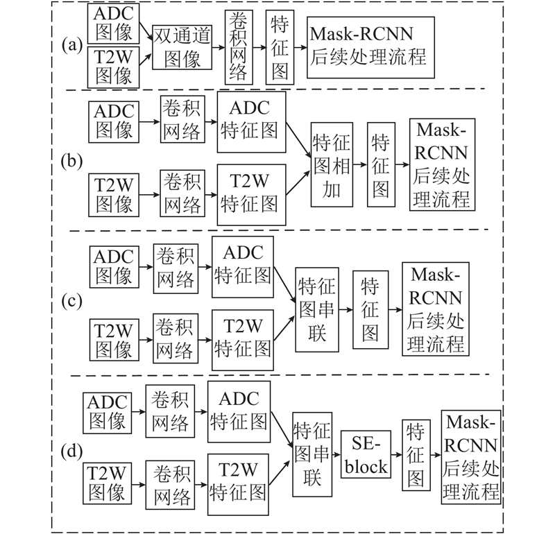
| SE-Mask-RCNN:多参数MRI前列腺癌分割方法 |
黄毅鹏1,2( ),胡冀苏1,2,钱旭升1,2,周志勇2,赵文露3,马麒3,沈钧康3,戴亚康2,*() ),胡冀苏1,2,钱旭升1,2,周志勇2,赵文露3,马麒3,沈钧康3,戴亚康2,*() |
1. 中国科学技术大学 生物医学工程学院(苏州),江苏 苏州 215163
2. 中国科学院 苏州生物医学工程技术研究所,江苏 苏州 215163
3. 苏州大学附属第二医院,江苏 苏州 215000 |
|
| SE-Mask-RCNN: segmentation method for prostate cancer on multi-parametric MRI |
| Yi-peng HUANG1,2(),Ji-su HU1,2,Xu-sheng QIAN1,2,Zhi-yong ZHOU2,Wen-lu ZHAO3,Qi MA3,Jun-kang SHEN3,Ya-kang DAI2,*() |
1. School of Biomedical Engineering (Suzhou), University of Science and Technology of China, Suzhou 215163, China
2. Suzhou Institute of Biomedical Engineering and Technology, Chinese Academy of Sciences, Suzhou 215163, China
3. The Second Affiliated Hospital of Suzhou University, Suzhou 215000, China |
引用本文:
黄毅鹏,胡冀苏,钱旭升,周志勇,赵文露,马麒,沈钧康,戴亚康. SE-Mask-RCNN:多参数MRI前列腺癌分割方法[J]. 浙江大学学报(工学版), 2021, 55(1): 203-212.
Yi-peng HUANG,Ji-su HU,Xu-sheng QIAN,Zhi-yong ZHOU,Wen-lu ZHAO,Qi MA,Jun-kang SHEN,Ya-kang DAI. SE-Mask-RCNN: segmentation method for prostate cancer on multi-parametric MRI. Journal of ZheJiang University (Engineering Science), 2021, 55(1): 203-212.
链接本文:
http://www.zjujournals.com/eng/CN/10.3785/j.issn.1008-973X.2021.01.024
或
http://www.zjujournals.com/eng/CN/Y2021/V55/I1/203
|
| 1 |
CHEN W, ZHENG R, BAADE P D, et al Cancer statistics in China, 2015[J]. CA: A Cancer Journal for Clinicians, 2016, 66 (2): 115- 132
doi: 10.3322/caac.21338
|
| 2 |
CHEN W, ZHENG R, ZENG H, et al Annual report on status of cancer in China, 2011[J]. Chinese Journal of Cancer Research, 2015, 27 (1): 2
|
| 3 |
陈金东 中国各类癌症的发病率和死亡率现状及发展趋势[J]. 遵义医学院学报, 2018, 41 (6): 653- 662
CHEN Jin-dong Trends of cancer incidence and mortality in China[J]. Journal of Zunyi Medical University, 2018, 41 (6): 653- 662
doi: 10.3969/j.issn.1000-2715.2018.06.001
|
| 4 |
王宁, 刘硕, 杨雷, 等 2018全球癌症统计报告解读[J]. 肿瘤综合治疗电子杂志, 2019, 5 (1): 87- 97
WANG Ning, LIU Shuo, YANG Lei, et al Interpretation on the report of global cancer statistics 2018[J]. Journal of Multidisciplinary Cancer Management (Electronic Version), 2019, 5 (1): 87- 97
|
| 5 |
SIEGEL R L, MILLER K D, JEMAL A Cancer statistics, 2019[J]. CA: A Cancer Journal for Clinicians, 2019, 69 (1): 7- 34
doi: 10.3322/caac.21551
|
| 6 |
PANEBIANCO V, BARCHETTI F, SCIARRA A, et al Multiparametric magnetic resonance imaging vs. standard care in men being evaluated for prostate cancer: a randomized study[J]. Urologic Oncology: Seminars and Original Investigations, 2015, 33 (1): 17.e1- 17.e7
doi: 10.1016/j.urolonc.2014.09.013
|
| 7 |
FüTTERER J J, BRIGANTI A, DE VISSCHERE P, et al Can clinically significant prostate cancer be detected with multiparametric magnetic resonance imaging? a systematic review of the literature[J]. European Urology, 2015, 68 (6): 1045- 1053
doi: 10.1016/j.eururo.2015.01.013
|
| 8 |
王建业, 陈鑫, 刘明, 等 MRI在前列腺癌诊断和治疗中的应用[J]. 磁共振成像, 2010, 1 (4): 253- 256
WANG Jian-ye, CHEN Xin, LIU Ming, et al MRI in clinical diagnosis and treatment of prostate cancer[J]. Chinese Journal of Magnetic Resonance Imaging, 2010, 1 (4): 253- 256
|
| 9 |
KOHL S, BONEKAMP D, SCHLEMMER H P, et al. Adversarial networks for the detection of aggressive prostate cancer [DB/OL]. [2017-02-26]. https://arxiv.org/abs/1702.08014.
|
| 10 |
蒋宏达, 叶西宁 一种改进的I-Unet网络的皮肤病图像分割算法[J]. 现代电子技术, 2019, 42 (12): 52- 56
JIANG Hong-da, YE Xi-ning An improved skin disease image segmentation algorithm based on I-Unet network[J]. Modern Electronics Technique, 2019, 42 (12): 52- 56
|
| 11 |
赖小波, 许茂盛, 徐小媚 多分类CNN的胶质母细胞瘤多模态MR图像分割[J]. 电子学报, 2019, 47 (8): 140- 149
LAI Xiao-bo, XU Mao-sheng, XU Xiao-mei Glioblastoma multiforme nulti-modal MRI images segmentation using multi-class CNN[J]. Acta Electronica Sinica, 2019, 47 (8): 140- 149
|
| 12 |
邢波涛, 李锵, 关欣 改进的全卷积神经网络的脑肿瘤图像分割[J]. 信号处理, 2018, 34 (8): 911- 922
XING Bo-tao, LI Qiang, GUAN Xin A brain tumor image segmentation method based on improved fully convolutional neural network[J]. Journal of Signal Processing, 2018, 34 (8): 911- 922
|
| 13 |
田萱, 王亮, 丁琪 基于深度学习的图像语义分割方法综述[J]. 软件学报, 2019, 30 (2): 250- 278
TIAN Xuan, WANG Liang, DING Qi Review of image semantic segmentation based on deep learning[J]. Journal of Software, 2019, 30 (2): 250- 278
|
| 14 |
RONNEBERGER O, FISCHER P, BROX T. U-net: convolutional networks for biomedical image segmentation [C] // International Conference on Medical Image Computing and Computer-Assisted Intervention. Munich: Springer, 2015: 234-241.
|
| 15 |
HE K, GKIOXARI G, DOLLáR P, et al. Mask R-CNN [C] // IEEE International Conference on Computer Vision. Washington D. C. : IEEE, 2017: 2961-2969.
|
| 16 |
TSEHAY Y K, LAY N S, ROTH H R, et al. Convolutional neural network based deep-learning architecture for prostate cancer detection on multi-parametric magnetic resonance images [C] // Medical Imaging 2017: Computer-Aided Diagnosis. Orlando: SPIE, 2017: 1013405.
|
| 17 |
YANG X, LIU C, WANG Z, et al Co-trained convolutional neural networks for automated detection of prostate cancer in multi-parametric MRI[J]. Medical Image Analysis, 2017, 42: 212- 227
doi: 10.1016/j.media.2017.08.006
|
| 18 |
YUAN Y, QIN W, BUYYOUNOUSKI M, et al Prostate cancer classification with multiparametric MRI transfer learning model[J]. Medical Physics, 2019, 46 (2): 756- 765
doi: 10.1002/mp.13367
|
| 19 |
HU J, SHEN L, SUN G. Squeeze-and-excitation networks [C] // Proceedings of the IEEE Conference on Computer Vision and Pattern Recognition. Salt Lake City: IEEE, 2018: 7132-7141.
|
| 20 |
LIU P, WANG S, TURKBEY B, et al. A prostate cancer computer-aided diagnosis system using multimodal magnetic resonance imaging and targeted biopsy labels [C] // Medical Imaging 2013: Computer-Aided Diagnosis. Lake Buena Vista: SPIE, 2013: 86701G.
|
| 21 |
YU Q, LIU F, SONG Y Z, et al. Sketch me that shoe [C]// Proceedings of the IEEE Conference on Computer Vision and Pattern Recognition. Las Vegas: IEEE, 2016: 799-807.
|
| 22 |
QIAN C, WANG L, GAO Y, et al In vivo MRI based prostate cancer localization with random forests and auto-context model[J]. Computerized Medical Imaging and Graphics, 2016, 52: 44- 57
doi: 10.1016/j.compmedimag.2016.02.001
|
| 23 |
MAZZETTI S, GIANNINI V, RUSSO F, et al Computer-aided diagnosis of prostate cancer using multi-parametric MRI: comparison between PUN and Tofts models[J]. Physics in Medicine and Biology, 2018, 63 (9): 095004
doi: 10.1088/1361-6560/aab956
|
| 24 |
MILLETARI F, NAVAB N, AHMADI S A. V-net: fully convolutional neural networks for volumetric medical image segmentation [C]// 2016 4th International Conference on 3D Vision. Stanford: IEEE, 2016: 565-571.
|
| 25 |
ISHIOKA J, MATSUOKA Y, UEHARA S, et al Computer‐aided diagnosis of prostate cancer on magnetic resonance imaging using a convolutional neural network algorithm[J]. BJU International, 2018, 122 (3): 411- 417
doi: 10.1111/bju.14397
|
| 26 |
陆洋, 柏根基 定量动态增强磁共振成像及其在前列腺癌诊断和治疗中的研究进展[J]. 磁共振成像, 2015, 6 (10): 796- 800
LU Yang, BO Gen-ji Progress of quantitative dynamic contrast-enhanced magnetic resonance imaging and in diagnosis and evaluation on the therapeutic response of prostate cancer[J]. Chinese Journal of Magnetic Resonance Imaging, 2015, 6 (10): 796- 800
doi: 10.3969/j.issn.1674-8034.2015.10.016
|
|
Viewed |
|
|
|
Full text
|
|
|
|
|
Abstract
|
|
|
|
|
Cited |
|
|
|
|
| |
Shared |
|
|
|
|
| |
Discussed |
|
|
|
|