|
|
|
| Medical image segmentation model based on KAN and CKAN optimization |
Shimeng LOU1( ),Yubin SHAO1,*(),Qingzhi DU1,Jingmin TANG1,Zetao ZHANG2 ),Yubin SHAO1,*(),Qingzhi DU1,Jingmin TANG1,Zetao ZHANG2 |
1. Faculty of Information Engineering and Automation, Kunming University of Science and Technology, Kunming 650500, China
2. Yunnan Province Key Laboratory for Media Integration, Kunming 650228, China |
|
|
|
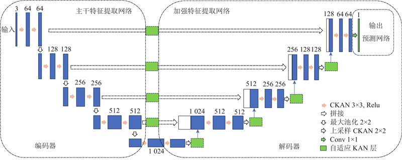
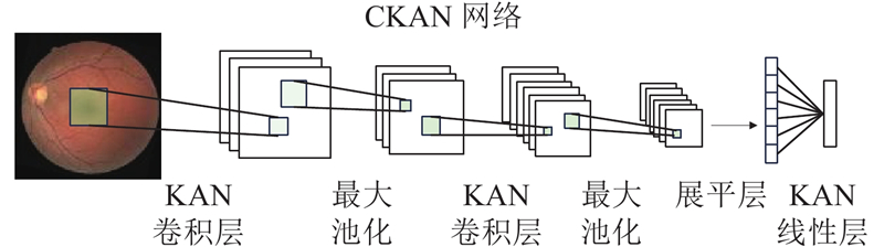
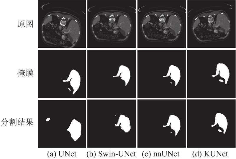
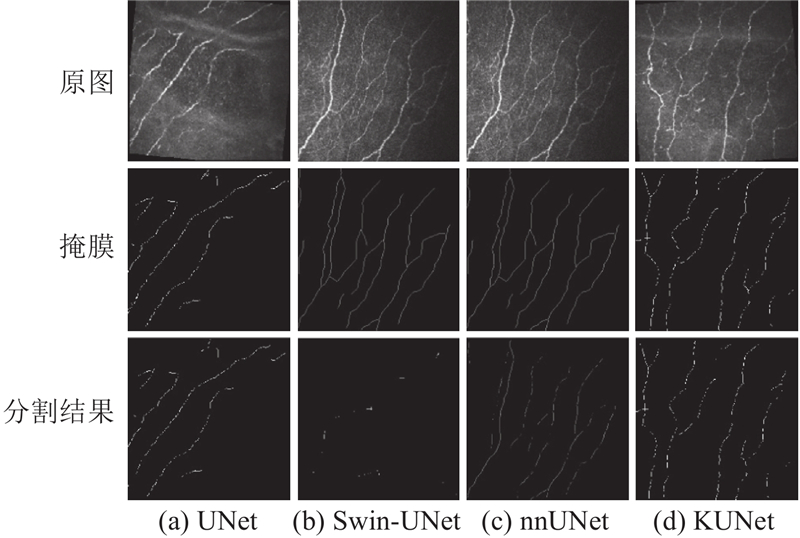
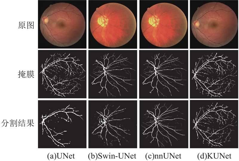
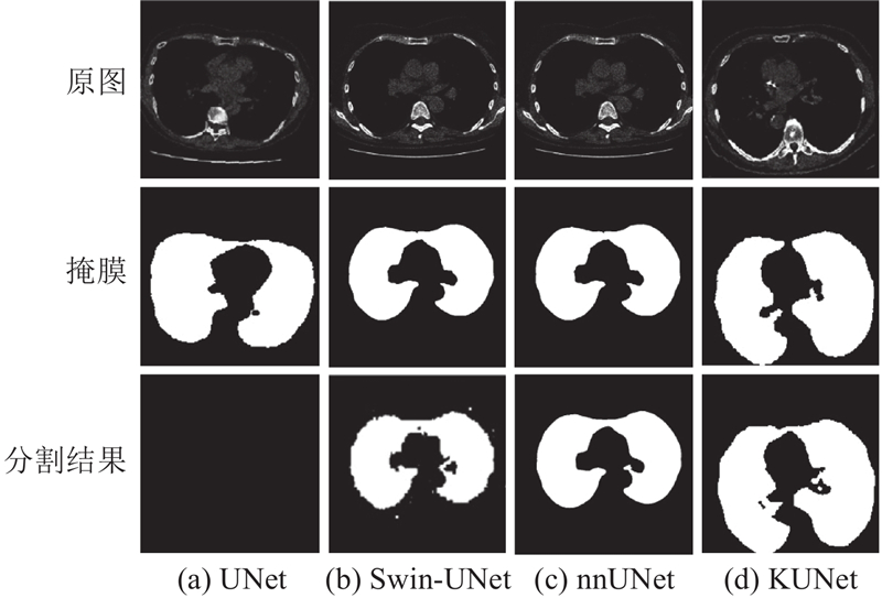
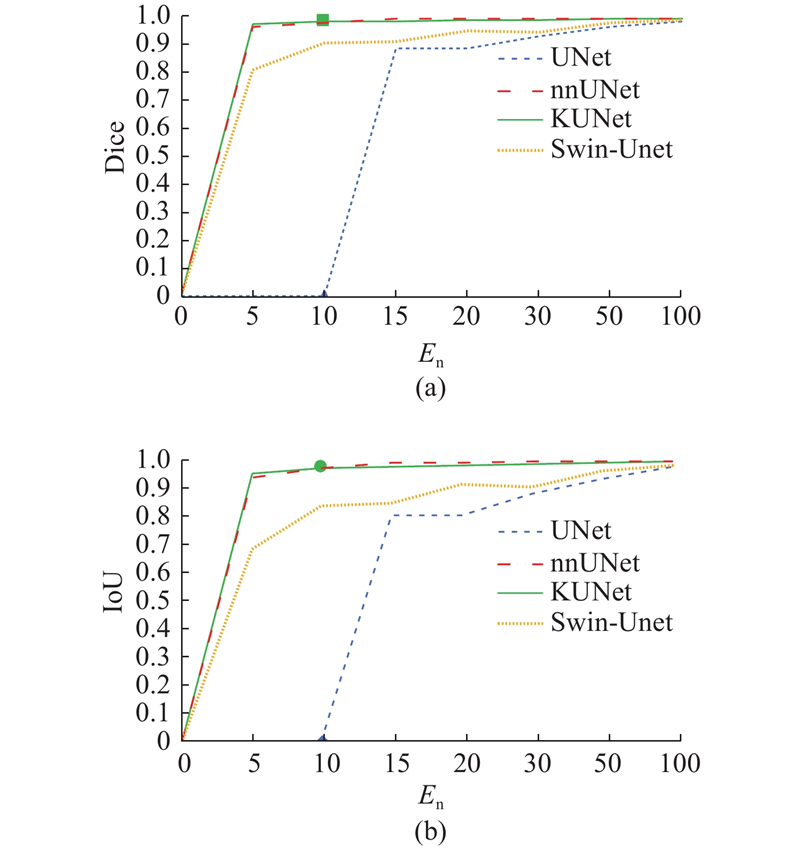
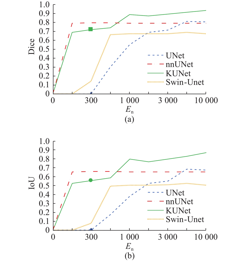
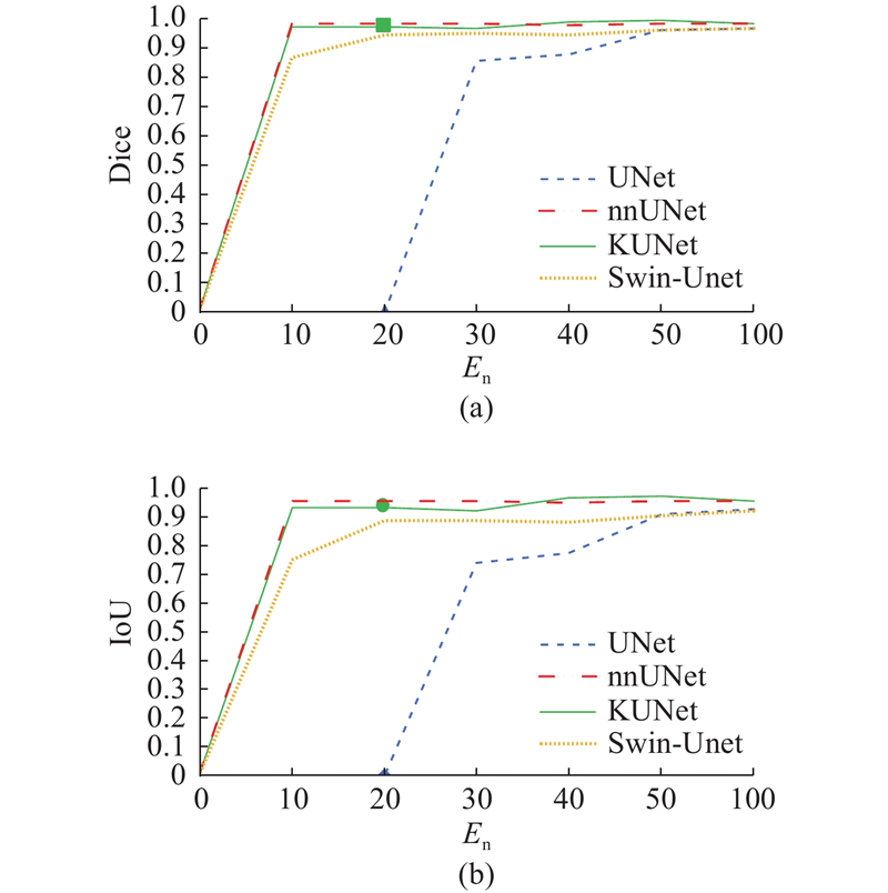
Abstract An optimized model KUNet based on Kolmogorov-Arnold network (KAN) and convolutional KAN (CKAN) was proposed to enhance the performance of the UNet model in order to address the limitation of the UNet model in complex feature extraction and generalization capability for medical image segmentation task. Traditional convolutional layer was replaced with CKAN, KAN feature enhancement module was introduced, and skip connection was optimized. Then the diversity and accuracy of feature extraction were improved while preserving structural information by incorporating an adaptive basis function learning mechanism. Comparative experiments were conducted against the UNet baseline model, nnUNet model and Swin-UNet model on four different multimodal datasets: LiTS, CORN, DRIVE and Lungs. Results showed that the average maximum absolute performance gap (MAPG) between the UNet baseline model and the KUNet model across the four datasets were 0.679 9 and 0.620 3 for Dice coefficient and IoU coefficient, respectively, and the KUNet model achieved average improvement metrics of 0.3213 and 0.2625 compared with the optimal or suboptimal model across the four datasets. The KUNet model was utilized to effectively extract more feature within short training cycle and improve the accuracy of image segmentation.
|
|
Received: 28 June 2025
Published: 06 May 2026
|
|
|
| Fund: 云南省媒体融合重点实验室资助项目(220245203). |
|
Corresponding Authors:
Yubin SHAO
E-mail: 2962772160@qq.com;shaoyubin999@qq.com
|
基于KAN与CKAN优化的医学图像分割模型
为了解决UNet模型在医学图像分割任务中的复杂特征提取与泛化能力不足的问题,提出基于Kolmogorov-Arnold网络(KAN)和卷积KAN(CKAN)的优化模型KUNet,增强UNet模型的性能. 通过用CKAN替换传统卷积层,引入KAN特征增强模块,优化跳跃连接,结合自适应基函数学习机制,在保留结构信息的同时提高特征提取的多样性与精度. 在4个不同的多模态数据集LiTS、CORN、DRIVE和Lungs上,与UNet基线模型、nnUNet模型和Swin-UNet模型进行对比实验. 结果表明,UNet基线模型与KUNet模型在4个数据集上Dice系数和IoU系数的平均最大性能差异指标(MAPG)分别为0.6799和0.6203,且KUNet模型相较于最优或次优模型在4个数据集上的平均提升指标为0.3213和0.2625. 利用KUNet模型,能够在短周期内有效提取到更多的特征,提升图像分割的准确度.
关键词:
图像分割,
UNet,
Kolmogorov-Arnold network(KAN),
卷积KAN(CKAN),
最大性能差异指标(MAPG)
|
|
| [1] |
GARCIA-GARCIA A, ORTS-ESCOLANO S, OPREA S, et al. A review on deep learning techniques applied to semantic segmentation [EB/OL]. [2025-08-17]. https://arxiv.org/abs/1704.06857.
|
|
|
| [2] |
MINAEE S, BOYKOV Y, PORIKLI F, et al Image segmentation using deep learning: a survey[J]. IEEE Transactions on Pattern Analysis and Machine Intelligence, 2021, 44 (7): 3523- 3542
|
|
|
| [3] |
ROTH H R, LU L, FARAG A, et al. DeepOrgan: multi-level deep convolutional networks for automated pancreas segmentation [C]//Medical Image Computing and Computer-Assisted Intervention. Cham: Springer, 2015: 556–564.
|
|
|
| [4] |
LITJENS G, KOOI T, BEJNORDI B E, et al A survey on deep learning in medical image analysis[J]. Medical Image Analysis, 2017, 42: 60- 88
doi: 10.1016/j.media.2017.07.005
|
|
|
| [5] |
DEVALLA S K, PHAM T H, PANDA S K, et al Towards label-free 3D segmentation of optical coherence tomography images of the optic nerve head using deep learning[J]. Biomedical Optics Express, 2020, 11 (11): 6356- 6378
doi: 10.1364/BOE.395934
|
|
|
| [6] |
CORDTS M, OMRAN M, RAMOS S, et al. The cityscapes dataset for semantic urban scene understanding [C]//Proceedings of the IEEE Conference on Computer Vision and Pattern Recognition. Las Vegas: IEEE, 2016: 3213–3223.
|
|
|
| [7] |
RONNEBERGER O, FISCHER P, BROX T. U-Net: convolutional networks for biomedical image segmentation [C]//Medical Image Computing and Computer-Assisted Intervention. Cham: Springer, 2015: 234–241.
|
|
|
| [8] |
ZHOU Z, RAHMAN SIDDIQUEE M M, TAJBAKHSH N, et al. UNet++: a nested U-Net architecture for medical image segmentation [C]//Deep Learning in Medical Image Analysis and Multimodal Learning for Clinical Decision Support. Cham: Springer, 2018: 3–11.
|
|
|
| [9] |
ZHAO H, SHI J, QI X, et al. Pyramid scene parsing network [C]//Proceedings of the IEEE Conference on Computer Vision and Pattern Recognition. Honolulu: IEEE, 2017: 6230–6239.
|
|
|
| [10] |
CHEN L C, PAPANDREOU G, KOKKINOS I, et al DeepLab: semantic image segmentation with deep convolutional nets, atrous convolution, and fully connected CRFs[J]. IEEE Transactions on Pattern Analysis and Machine Intelligence, 2018, 40 (4): 834- 848
doi: 10.1109/TPAMI.2017.2699184
|
|
|
| [11] |
CAO H, WANG Y, CHEN J, et al. Swin-Unet: Unet-like pure transformer for medical image segmentation [C]// European Conference on Computer Vision. Cham: Springer, 2023: 205–218.
|
|
|
| [12] |
ISENSEE F, JAEGER P F, KOHL S A A, et al nnU-NET: a self-configuring method for deep learning-based biomedical image segmentation[J]. Nature Methods, 2021, 18 (2): 203- 211
doi: 10.1038/s41592-020-01008-z
|
|
|
| [13] |
ISENSEE F, PETERSEN J, KLEIN A, et al. nnU-NET: self-adapting framework for U-Net-based medical image segmentation [EB/OL]. [2025-08-17]. https://arxiv.org/abs/1809.10486.
|
|
|
| [14] |
MOU L, ZHAO Y, CHEN L, et al. CS-Net: channel and spatial attention network for curvilinear structure segmentation [C]//Medical Image Computing and Computer Assisted Intervention. Cham: Springer, 2019: 721–730.
|
|
|
| [15] |
STAAL J, ABRAMOFF M D, NIEMEIJER M, et al Ridge-based vessel segmentation in color images of the retina[J]. IEEE Transactions on Medical Imaging, 2004, 23 (4): 501- 509
doi: 10.1109/TMI.2004.825627
|
|
|
| [16] |
LIU Z, WANG Y, VAIDYA S, et al. KAN: Kolmogorov-Arnold networks [EB/OL]. [2025-08-17]. https://arxiv.org/abs/2404.19756.
|
|
|
| [17] |
BODNER A D, TEPSICH A S, SPOLSKI J N, et al. Convolutional Kolmogorov-Arnold networks [EB/OL]. [2025-08-17]. https://arxiv.org/abs/2406.13155.
|
|
|
| [18] |
LI C, LIU X, LI W, et al U-KAN makes strong backbone for medical image segmentation and generation[J]. Proceedings of the AAAI Conference on Artificial Intelligence, 2025, 39 (5): 4652- 4660
doi: 10.1609/aaai.v39i5.32491
|
|
|
| [19] |
MA X, WANG Z, HU Y, et al. Kolmogorov-Arnold network for remote sensing image semantic segmentation [EB/OL]. [2025-08-17]. https://arxiv.org/abs/2501.07390.
|
|
|
| [20] |
AGRAWAL A, AGRAWAL A, GUPTA S, et al. KAN-Mamba FusionNet: redefining medical image segmentation with non-linear modeling [EB/OL]. [2025-08-17]. https://arxiv.org/abs/2411.11926.
|
|
|
| [21] |
OKTAY O, SCHLEMPER J, FOLGOC L L, et al. Attention U-Net: learning where to look for the pancreas [EB/OL]. [2025-08-17]. https://arxiv.org/abs/1804.03999.
|
|
|
| [22] |
ZHANG Z, LIU Q, WANG Y Road extraction by deep residual U-Net[J]. IEEE Geoscience and Remote Sensing Letters, 2018, 15 (5): 749- 753
doi: 10.1109/LGRS.2018.2802944
|
|
|
| [23] |
BILIC P, CHRIST P, LI H B, et al The Liver tumor segmentation benchmark (LiTS)[J]. Medical Image Analysis, 2023, 84: 102680
doi: 10.1016/j.media.2022.102680
|
|
|
| [24] |
SCHOENBERG I J. Contributions to the problem of approximation of equidistant data by analytic functions: part A. -on the problem of smoothing or graduation. a first class of analytic approximation formulae [J]. Quarterly of Applied Mathematics, 1946, 4(1): 45–99.
|
|
|
| [25] |
HOLLADAY J C A smoothest curve approximation[J]. Mathematical Tables and Other Aids to Computation, 1957, 11 (60): 233- 243
doi: 10.1090/s0025-5718-1957-0093894-6
|
|
|
| [26] |
MILLETARI F, NAVAB N, AHMADI S A. V-net: fully convolutional neural networks for volumetric medical image segmentation [C]// Proceedings of the Fourth International Conference on 3D Vision. Stanford: IEEE, 2016: 565–571.
|
|
|
| [27] |
EVERINGHAM M, VAN GOOL L, WILLIAMS C K I, et al The pascal visual object classes (VOC) challenge[J]. International Journal of Computer Vision, 2010, 88 (2): 303- 338
doi: 10.1007/s11263-009-0275-4
|
|
|
| [28] |
TAGHANAKI S A, ZHENG Y, ZHOU K S, et al Combo loss: handling input and output imbalance in multi-organ segmentation[J]. Computerized Medical Imaging and Graphics, 2019, 75: 24- 33
doi: 10.1016/j.compmedimag.2019.04.005
|
|
|
|
Viewed |
|
|
|
Full text
|
|
|
|
|
Abstract
|
|
|
|
|
Cited |
|
|
|
|
| |
Shared |
|
|
|
|
| |
Discussed |
|
|
|
|