| Mechanical and Energy Engineering |
|


|
|
|
| Classification on histological subtypes of lung adenocarcinoma from low-resolution CT images based on DenseNet |
Jing YANG1,2( ),Chen GENG2,Hai-lin WANG3,Jian-song JI3,Ya-kang DAI2,*() ),Chen GENG2,Hai-lin WANG3,Jian-song JI3,Ya-kang DAI2,*() |
1. University of Science and Technology of China, Hefei 230026, China
2. Suzhou Institute of Biomedical Engineering and Technology, Chinese Academy of Sciences, Suzhou 215163, China
3. The Central Hospital of Lishui City, Lishui 323000, China |
|
|
|
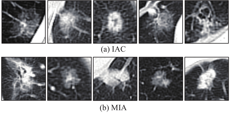
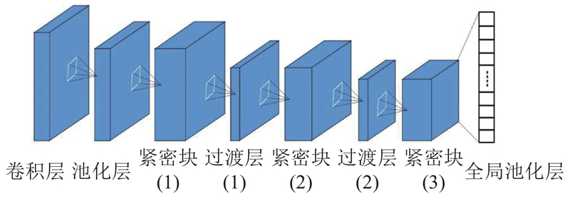
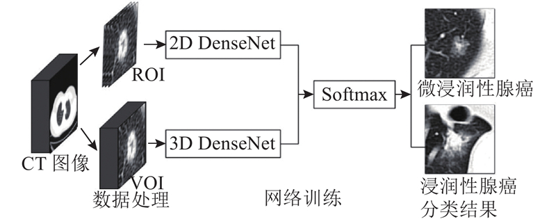
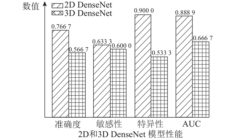
Abstract A deep learning method based on DenseNet was proposed to distinguish between IAC and MIA from mixed ground glass nodules low-resolution CT images with 5 mm slice thickness, in order to classify histological subtypes of lung adenocarcinoma from low-dose CT images with low resolution. Samples were obtained from 105 low-resolution CT images with 5 mm slice thickness of 105 patients in the Central Hospital of Lishui City. The data was divided into training set and testing set. Then the training set was augmented; 2D and 3D DenseNet deep learning models were built to distinguish between IAC and MIA. The accuracy, sensitivity, specificity and the area under the receiver operating characteristic curve of the proposed 2D DenseNet method achieved 76.67%, 63.33%, 90.00% and 0.888 9, respectively, which was better than 3D DenseNet and other deep learning models. The deep learning method, especially the 2D DenseNet, may assist doctors in lung cancer screening to predict and guide histological subtypes of patients, which can quickly provide more accurate diagnosis results even under condition of low image resolution.
|
|
Received: 05 January 2019
Published: 22 May 2019
|
|
|
|
Corresponding Authors:
Ya-kang DAI
E-mail: 18366136246@163.com;daiyk@sibet.ac.cn
|
基于DenseNet的低分辨CT影像肺腺癌组织学亚型分类
为了实现在低剂量、低分辨率CT扫描影像中对肺腺癌组织学亚型的分类鉴别,提出一种基于DenseNet的深度学习方法,从混合性磨玻璃结节(mGGNs)5 mm层厚的低分辨率CT影像中预测IAC和MIA病理分类. 从丽水市中心医院105例患者的105个5 mm层厚低分辨率CT图像中选取样本,划分训练集和测试集后,对训练集进行数据扩展,构建深度学习2D和3D DenseNet模型,分类鉴别IAC和MIA. 2D DenseNet模型的分类准确度为76.67%,敏感性为63.33%,特异性为90.00%,受试者工作特征曲线下的区域面积为0.888 9,显著优于3D DenseNet模型和其他几种深度学习网络模型. 深度学习技术,尤其是2D DenseNet模型,可辅助并指导医生在肺癌CT筛查中对患者的肺腺癌组织学亚型进行预判,特别是在图像分辨率较低的情况下,仍能够快速提供较为准确的诊断.
关键词:
深度学习,
DenseNet,
混合性磨玻璃结节,
肺腺癌,
厚层CT
|
|
| [1] |
SIEGEL R L, MILLER K D, JEMAL A Cancer statistics, 2018[J]. CA: a Cancer Journal for Clinicians, 2018, 68 (1): 7- 30
doi: 10.3322/caac.21442
|
|
|
| [2] |
YANG J, WANG H, GENG C, et al Advances in intelligent diagnosis methods for pulmonary ground-glass opacity nodules[J]. Biomedical Engineering Online, 2018, 17 (1): 20
doi: 10.1186/s12938-018-0435-2
|
|
|
| [3] |
HENSCHKE C I, YANKELEVITZ D F, MIRTCHEVA R, et al CT screening for lung cancer: frequency and significance of part-solid and nonsolid nodules[J]. AJR American Journal of Roentgenology, 2002, 178 (5): 1053- 1057
doi: 10.2214/ajr.178.5.1781053
|
|
|
| [4] |
COHEN J G, REYMOND E, MEDICI M, et al CT-texture analysis of subsolid nodules for differentiating invasive from in-situ and minimally invasive lung adenocarcinoma subtypes[J]. Diagnostic and Interventional Imaging, 2018, 99 (5): 291- 299
doi: 10.1016/j.diii.2017.12.013
|
|
|
| [5] |
TRAVIS W D, BRAMBILLA E, NOGUCHI M, et al International association for the study of lung cancer/American thoracic society/European respiratory society international multidisciplinary classification of lung adenocarcinoma[J]. Journal of Thoracic Oncology Official Publication of the International Association for the Study of Lung Cancer, 2011, 6 (2): 244- 285
doi: 10.1097/JTO.0b013e318206a221
|
|
|
| [6] |
TRAVIS W D, BRAMBILLA E, NICHOLSON A G, et al The 2015 world health organization classification of lung tumors: impact of genetic, clinical and radiologic advances since the 2004 classification[J]. Journal of Thoracic Oncology Official Publication of the International Association for the Study of Lung Cancer, 2015, 10 (9): 1243- 1260
doi: 10.1097/JTO.0000000000000630
|
|
|
| [7] |
NAIDICH D P, BANKIER A A, MACMAHON H, et al Recommendations for the management of subsolid pulmonary nodules detected at CT: a statement from the fleischner society[J]. Radiology, 2013, 266 (1): 304- 317
doi: 10.1148/radiol.12120628
|
|
|
| [8] |
YUE X, LIU S, LIU S, et al HRCT morphological characteristics distinguishing minimally invasive pulmonary adenocarcinoma from invasive pulmonary adenocarcinoma appearing as subsolid nodules with a diameter of </=3 cm[J]. Clinical Radiology, 2018, 73 (4): 411. e7- 411. e15
doi: 10.1016/j.crad.2017.11.014
|
|
|
| [9] |
VAN SCHIL P E, ASAMURA H, RUSCH V W, et al Surgical implications of the new IASLC/ATS/ERS adenocarcinoma classification[J]. European Respiratory Journal, 2012, 39 (2): 478- 486
doi: 10.1183/09031936.00027511
|
|
|
| [10] |
LIU S, WANG R, ZHANG Y, et al Precise diagnosis of intraoperative frozen section is an effective method to guide resection strategy for peripheral small-sized lung adenocarcinoma[J]. Journal of Clinical Oncology, 2016, 34 (4): 307- 313
doi: 10.1200/JCO.2015.63.4907
|
|
|
| [11] |
ZHANG J, WU J, TAN Q, et al Why do pathological stage IA lung adenocarcinomas vary from prognosis?: a clinicopathologic study of 176 patients with pathological stage IA lung adenocarcinoma based on the IASLC/ATS/ERS classification[J]. Journal of Thoracic Oncology Official Publication of the International Association for the Study of Lung Cancer, 2013, 8 (9): 1196- 1202
doi: 10.1097/JTO.0b013e31829f09a7
|
|
|
| [12] |
涂文婷, 范丽, 顾亚峰, 等 计算机辅助定量分析对磨玻璃密度型肺腺癌浸润性的诊断价值[J]. 临床放射学杂志, 2018, 37 (3): 497- 502
TU Wen-ting, FAN Li, GU Ya-feng, et al The value of computer-aided quantitative analysis in the diagnosis of invasiveness of lung adenocarcinoma manifesting as ground glass nodule[J]. Journal of Clinical Radiology, 2018, 37 (3): 497- 502
|
|
|
| [13] |
SON J Y, LEE H Y, LEE K S, et al Quantitative CT analysis of pulmonary ground-glass opacity nodules for the distinction of invasive adenocarcinoma from pre-invasive or minimally invasive adenocarcinoma[J]. PLoS One, 2014, 9 (8): e104066
doi: 10.1371/journal.pone.0104066
|
|
|
| [14] |
左玉强, 冯平勇, 孟庆春, 等 肺纯磨玻璃结节微浸润腺癌与浸润性腺癌的CT鉴别诊断[J]. 临床放射学杂志, 2017, 36 (4): 495- 498
ZUO Yu-qiang, FENG Ping-yong, MENG Qing-chun, et al CT differential diagnoses of pulmonary minimally invasive adenocarcinoma and invasive adenocarcinoma presenting as pure ground glass nodule[J]. Journal of Clinical Radiology, 2017, 36 (4): 495- 498
|
|
|
| [15] |
WANG H, ZHAO T, LI L C, et al A hybrid CNN feature model for pulmonary nodule malignancy risk differentiation[J]. Journal of X-ray Science and Technology, 2018, 26 (2): 171- 187
doi: 10.3233/XST-17302
|
|
|
| [16] |
ZHAO X, LIU L, QI S, et al Agile convolutional neural network for pulmonary nodule classification using CT images[J]. International Journal of Computer Assisted Radiology and Surgery, 2018, 13 (4): 585- 595
doi: 10.1007/s11548-017-1696-0
|
|
|
| [17] |
HUANG G, LIU Z, MAATEN L V D, et al. Densely connected convolutional networks [C] // 2017 IEEE Conference on Computer Vision and Pattern Recognition. Hawaii: IEEE, 2017: 2261–2269.
|
|
|
| [18] |
SRIVASTAVA N, HINTON G, KRIZHEVSKY A, et al Dropout: a simple way to prevent neural networks from overfitting[J]. Journal of Machine Learning Research, 2014, 15 (1): 1929- 1958
|
|
|
| [19] |
BAHRAMPOUR S, RAMAKRISHNAN N, SCHOTT L, et al Comparative study of deep learning software frameworks[J]. Computer Science, 2016,
|
|
|
| [20] |
LING C X, HUANG J, ZHANG H AUC: a better measure than accuracy in comparing learning algorithms[J]. Lecture Notes in Computer Science, 2003, 329- 341
|
|
|
| [21] |
LECUN Y, BOTTOU L, BENGIO Y, et al Gradient-based learning applied to document recognition[J]. P Ieee, 1998, 86 (11): 2278- 2324
doi: 10.1109/5.726791
|
|
|
| [22] |
KRIZHEVSKY A, SUTSKEVER I, HINTON G E ImageNet classification with deep convolutional neural networks[J]. Commun ACM, 2017, 60 (6): 84- 90
doi: 10.1145/3098997
|
|
|
| [23] |
HE K, ZHANG X, REN S, et al. Deep residual learning for image recognition [C] // 2016 IEEE Conference on Computer Vision and Pattern Recognition. Seattle: IEEE, 2016: 770–778.
|
|
|
|
Viewed |
|
|
|
Full text
|
|
|
|
|
Abstract
|
|
|
|
|
Cited |
|
|
|
|
| |
Shared |
|
|
|
|
| |
Discussed |
|
|
|
|