|
|
|
| Breast cancer histopathological image classification using multi-scale channel squeeze-and-excitation model |
Tao MING1,Dan WANG2,Ji-chang GUO1,*( ),Qiang LI3 ),Qiang LI3 |
1. School of Electrical and Information Engineering, Tianjin University, Tianjin 300072, China
2. Department of Pathology, General Hospital, Tianjin Medical University, Tianjin 300052, China
3. School of Microelectronics, Tianjin University, Tianjin 300072, China |
|
|
|
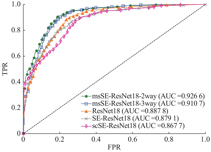
Abstract A deep learning-based classification algorithm was proposed aiming at the problem of automatic classification algorithms focusing on breast cancer histopathological images. Channel squeeze-and-excitation (SE) model is an attention model applied to the feature channels. Useless features can be suppressed with learned channel weights so as to recalibrate the feature channels for better classification accuracy. A multi-scale channel SE model was proposed, and a convolutional neural network named msSE-ResNet was designed in order to make the result of channel recalibration more accurate. Multi-scale features were obtained by max-pooling layers and served as inputs to subsequent channel SE models, and the result of channel recalibration was improved by merging channel weights learned under different feature scales. Experiments were conducted on public dataset BreaKHis. Results show that the network can reach an accuracy of 88.87% on the task of classifying benign/malignant breast histopathological images, and can remain good robustness to histopathological images acquired under different magnifications.
|
|
Received: 18 June 2019
Published: 05 July 2020
|
|
|
|
Corresponding Authors:
Ji-chang GUO
E-mail: jcguo@tju.edu.cn
|
基于多尺度通道重校准的乳腺癌病理图像分类
针对乳腺癌病理图像的自动分类问题,提出基于深度学习的分类算法. 通道重校准模型是作用于特征通道的注意力模型,可以利用学习到的通道权重对无用特征进行抑制来实现对特征通道的重校准,以达到更高的分类准确率. 为了使通道重校准的结果更加准确,提出多尺度通道重校准模型,设计卷积神经网络 msSE-ResNet. 多尺度特征通过网络中的最大池化层获得并作为后续通道重校准模型的输入,将不同尺度下学到的通道权重进行融合,可以改善通道重校准的结果. 该实验在公开数据集BreaKHis上开展. 实验结果表明,该网络对良性/恶性乳腺病理图像分类任务达到88.87%的分类精度,可以对不同放大倍数下获取的病理图像具有较好的鲁棒性.
关键词:
乳腺癌病理图像分类,
深度学习,
残差网络,
多尺度特征,
通道重校准模型
|
|
| [1] |
FAN L, STRASSER-WEIPPL K, LI J J, et al Breast cancer in China[J]. Lancet Oncology, 2014, 15 (7): 279- 289
doi: 10.1016/S1470-2045(13)70567-9
|
|
|
| [2] |
LEONG A S-Y, ZHUANG Z P The changing role of pathology in breast cancer diagnosis and treatment[J]. Pathobiology, 2011, 78: 99- 114
doi: 10.1159/000292644
|
|
|
| [3] |
VETA M, PLUIM J P, VAN DIEST P J, et al Breast cancer histopathology image analysis: a review[J]. IEEE Transactions on Biomedical Engineering, 2014, 61 (5): 1400- 1411
doi: 10.1109/TBME.2014.2303852
|
|
|
| [4] |
SPANHOL F A, OLIVEIRA L S, PETITJEAN C, et al A dataset for breast cancer histopathological image classification[J]. IEEE Transactions on Biomedical Engineering, 2016, 63 (7): 1455- 1462
doi: 10.1109/TBME.2015.2496264
|
|
|
| [5] |
GUPTA V, BHAVSAR A. Breast cancer histopathological image classification: is magnification important? [C] // Proceedings of IEEE Conference on Computer Vision and Pattern Recognition Workshops. Honolulu, USA: IEEE, 2017: 769-776.
|
|
|
| [6] |
CIRESAN D C, GIUSTI A, GAMBARDELLA L M, et al. Mitosis detection in breast cancer histology images with deep neural networks [C] // Proceedings of Medical Image Computing and Computer-Assisted Intervention. Berlin, German: Springer, 2013: 411-418.
|
|
|
| [7] |
ARAúJO T, ARESTA G, CASTRO E, et al Classification of breast cancer histology images using convolutional neural networks[J]. PLos One, 2017, 12 (6): e0177544
doi: 10.1371/journal.pone.0177544
|
|
|
| [8] |
SPANHOL F A, OLIVEIRA L S, PETITJEAN C, et al. Breast cancer histopathological image classification using convolutional neural networks [C] // Proceedings of International Joint Conference on Neural Networks. Vancouver, Canada: IEEE, 2016: 2560-2567.
|
|
|
| [9] |
BAYRAMOGLU N, KANNALA J, HEIKKIL? J. Deep learning for magnification independent breast cancer histopathology image classification [C] // Proceedings of International Conference on Pattern Recognition. Cancun, Mexico: IEEE, 2016: 2441-2446.
|
|
|
| [10] |
SONG Y, ZOU J J, CHANG H, et al. Adapting Fisher vectors for histopathology image classification [C] // Proceedings of the IEEE 14th International Symposium on Biomedical Imaging. Melbourne: IEEE, 2017: 600-603.
|
|
|
| [11] |
HU J, SHEN L, SUN G. Squeeze-and-Excitation network [C] // Proceedings of the IEEE Conference on Computer Vision and Pattern Recognition. Salt Lake City: IEEE, 2018: 7132-7141.
|
|
|
| [12] |
HE K, ZHANG X, REN S, et al. Deep residual learning for image recognition [C] // Proceedings of the IEEE Conference on Computer Vision and Pattern Recognition. Las Vegas, USA: IEEE, 2016: 770-778.
|
|
|
| [13] |
BAHDANAU D, CHO K, BENGIO Y. Neural machine translation by jointly learning to align and translate [EB/OL]. [2019–03–01]. https://arxiv.org/abs/1409.0473.
|
|
|
| [14] |
VASWANI A, SHAZEER N, PARMAR N, et al. Attention is all you need [C]// Proceedings of Neural Information Processing Systems. Long Beach, USA: Curran Associates, Inc., 2017: 5998-6008.
|
|
|
| [15] |
WANG F, JIANG M, QIAN C, et al. Residual attention network for image classification [C] // Proceedings of IEEE Conference on Computer Vision and Pattern Recognition. Honolulu, USA: IEEE, 2017: 6450-6458.
|
|
|
| [16] |
ZHU Y Y, WANG J, XIE L X, et al. Attention-based pyramid aggregation network for visual place recognition [C] // Proceedings of International Conference on Multimedia. Seoul, Korea: ACM, 2018: 99-107.
|
|
|
| [17] |
SIMONYAN K, ZISSERMAN A. Very deep convolutional networks for large-scale image recognition [EB/OL]. 2019–04–23. https://arxiv.org/abs/1409.1556.
|
|
|
| [18] |
SZEGEDY C, LIU W, JIA Y, et al. Going deeper with convolutions [C] // Proceedings of IEEE Conference on Computer Vision and Pattern Recognition. Boston, USA: IEEE, 2015: 1-9.
|
|
|
| [19] |
NAIR V, HINTON G E. rectified linear units improve restricted Boltzmann machine [C] // International Conference on International Conference on Machine Learning. Haifa, Israel: Omnipress, 2010: 807-814.
|
|
|
| [20] |
HE K, ZHANG X, REN S, et al Spatial pyramid pooling in deep convolutional networks for visual recognition[J]. IEEE Transactions on Pattern Analysis and Machine Intelligence, 2015, 37 (9): 1904- 1916
doi: 10.1109/TPAMI.2015.2389824
|
|
|
| [21] |
LIU W, ANGUELOV D, ERHAN D, et al. SSD: single shot multibox detector [C] // Proceedings of European Conference on Computer Vision. Amsterdam: Springer, 2016: 21-37.
|
|
|
| [22] |
LIN T, DOLLAR P, GIRSHICK R. Feature pyramid networks for object detection [C] // Proceedings of IEEE Conference on Computer Vision and Pattern Recognition. Honolulu: IEEE, 2017: 2117-2125.
|
|
|
| [23] |
ZHAO H, SHI J, QI X, et el. Pyramid scene parsing network [C] // Proceedings of IEEE Conference on Computer Vision and Pattern Recognition. Honolulu: IEEE, 2017: 6230-6239.
|
|
|
| [24] |
ZHAO H, QI X, SHEN X, et al. ICNet for real-time semantic segmentation on high-resolution images [C] // Proceedings of European Conference on Computer Vision. Munich, Germany: Springer, 2018: 418-434.
|
|
|
| [25] |
KAMNITASA K, LEDIG C, NEWCOMBE V F, et al Efficient multi-scale 3D CNN with fully connected CRF for accurate brain lesion segmentation[J]. Medical Image Analysis, 2017, 36: 61- 78
doi: 10.1016/j.media.2016.10.004
|
|
|
| [26] |
PASZKE A, GROSS S, MASSA F, et al. PyTorch: an imperative style, high-performing deep learning library [C] // Proceedings of Neural Information Processing Systems. Vancouver: Curran Associates, Inc., 2019: 8024-8035.
|
|
|
|
Viewed |
|
|
|
Full text
|
|
|
|
|
Abstract
|
|
|
|
|
Cited |
|
|
|
|
| |
Shared |
|
|
|
|
| |
Discussed |
|
|
|
|