| Computer Technology and Image Processing |
|


|
|
|
| Deep segmentation method of tumor boundaries from MR images of patients with nasopharyngeal carcinoma using multi-modality and multi-dimension fusion |
Yan-jia HONG1( ),Tie-bao MENG2,Hao-jiang LI2,Li-zhi LIU2,Li LI2,Shuo-yu XU2,Sheng-wen GUO1,*() ),Tie-bao MENG2,Hao-jiang LI2,Li-zhi LIU2,Li LI2,Shuo-yu XU2,Sheng-wen GUO1,*() |
1. Department of Biomedical Engineering, South China University of Technology, Guangzhou 510006, China
2. Medical Image Center, Sun Yat-sen University Cancer Center, Guangzhou 510060, China |
|
|
|
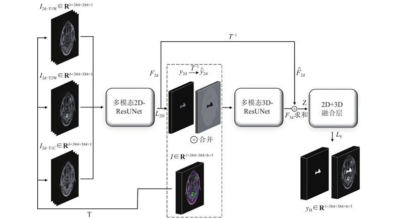
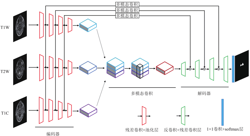
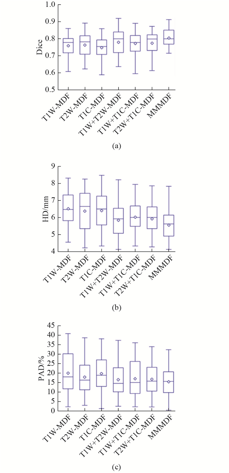
Abstract First, T1-weighted (T1W), T2-weighted (T2W) and T1 enhanced structural MR images of 421 patients were collected, the tumor boundaries of all images were delineated manually by two experienced doctors as the ground truth, the images and ground truth of 346 patients were considered as training set and the remaining images and corresponding ground truth of 75 patients were selected as independent testing set. Second, three single modality, based multi-dimension deep convolutional neural networks (CNN) and three two-modality multi-dimension fusion deep convolutional networks and a multi-modality multi-dimension fusion (MMMDF) deep convolutional neural network were constructed, and the networks were trained and tested, respectively. Finally, the performance of the three methods were evaluated by using three indexes, including Dice, Hausdorff distance (HD) and percentage area difference (PAD). The experimental results show that the MMMDF CNNs can acquire the best performances, followed by the two-modality multi-dimental fusion CNNs, while the single modlity multi-dimension CNNs achieves the worst measures.. This study demonstrates that the MMMDF-CNN combining multi-modality images and incorporating 2D with 3D images features can effectively fulfill accurate segmentation on tumors of MR images from NPC patients.
|
|
Received: 02 March 2019
Published: 05 March 2020
|
|
|
|
Corresponding Authors:
Sheng-wen GUO
E-mail: 531679559@qq.com;shwguo@scut.edu.cn
|
多模态多维信息融合的鼻咽癌MR图像肿瘤深度分割方法
收集421名鼻咽癌患者头颈部水平位T1加权(T1W)、T2加权(T2W)以及T1增强(T1C)三种模态MR图像,并由2名经验丰富的临床医生对图像中的肿瘤区域进行勾画,将其中346位患者的多模态图像及其标签作为训练集,将剩余75位患者的多模态图像及其标签作为独立测试集;分别构建单模态多维信息融合、两模态多维信息融合以及多模态多维信息融合(MMMDF)的卷积神经网络(CNN),并对模型进行训练和测试;使用Dice、豪斯多夫距离(HD)与面积差占比(PAD)评估3种模型的性能,结果表明,多模态多维融合模型的性能最优,两模态多维信息融合模型性能次之,单模态多维信息融合模型性能最差. 结果证明,多模态二维与三维特征融合的深度卷积网络能够准确有效地分割鼻咽癌MR图像中的肿瘤.
关键词:
鼻咽癌,
MR图像,
分割,
多模态多维度,
深度学习
|
|
| [1] |
CHANG E T, ADAMI H O The enigmatic epidemiology of nasopharyngeal carcinoma[J]. Cancer Epidemiology and Prevention Biomarkers, 2006, 15 (10): 1765- 1777
doi: 10.1158/1055-9965.EPI-06-0353
|
|
|
| [2] |
STEWART B W, WILD C. World cancer report 2014 [M]. Lyon: International Agency for Research on Cancer , 2014.
|
|
|
| [3] |
邓伟, 黄天壬, 陈万青, 等 中国2003—2007年鼻咽癌发病与死亡分析[J]. 肿瘤, 2012, 32 (3): 189- 193
DENG Wei, HUANG Tian-Ren, CHEN Wan-Qing, et al Analysis of the incidence and mortality of nasopharyngeal carcinoma in China from 2003 to 2007[J]. Tumor, 2012, 32 (3): 189- 193
doi: 10.3781/j.issn.1000-7431.2012.03.007
|
|
|
| [4] |
HUANG K W, ZHAO Z Y, GONG Q, et al. Nasopharyngeal carcinoma segmentation via HMRF-EM with maximum entropy [C] // 2015 37th Annual International Conference of the IEEE Engineering in Medicine and Biology Society (EMBC). Milan: IEEE, 2015: 2968-2972.
|
|
|
| [5] |
RITTHIPRAVAT P, TATANUM C, BHONGMAKAPAT T, et al. Automatic segmentation of nasopharyngeal carcinoma from CT images [C] // 2008 International Conference on BioMedical Engineering and Informatics. Sanya: IEEE Computer Society, 2008, 2: 18-22.
|
|
|
| [6] |
ZHOU J, CHAN K L, XU P, et al. Nasopharyngeal carcinoma lesion segmentation from MR images by support vector machine [C] // 3rd IEEE International Symposium on Biomedical Imaging: Nano to Macro, 2006. Arlington: IEEE, 2006: 1364-1367.
|
|
|
| [7] |
MOHAMMED M A, GHANI M K A, HAMED R I, et al Artificial neural networks for automatic segmentation and identification of nasopharyngeal carcinoma[J]. Journal of Computer Science, 2017, 21: 263- 274
doi: 10.1016/j.jocs.2017.03.026
|
|
|
| [8] |
MEN K, CHEN X, ZHANG Y, et al Deep deconvolutional neural network for Target segmentation of nasopharyngeal cancer in Planning computed Tomography images[J]. Frontiers in Oncology, 2017, 7: 315
doi: 10.3389/fonc.2017.00315
|
|
|
| [9] |
SIMONYAN K, ZISSERMAN A. Very deep convolutional networks for large-scale image recognition[J]. arXiv Preprint arXiv: 1409.1556, 2014.
|
|
|
| [10] |
LI Q L, XU Y, CHEN Z, et al Tumor segmentation in contrast-enhanced magnetic resonance imaging for nasopharyngeal carcinoma: deep learning with convolutional neural network[J]. BioMed Research International, 2018, 2018, 5: 1- 7
|
|
|
| [11] |
MA Z Q, WU X, SONG Q, et al Automated nasopharyngeal carcinoma segmentation in magnetic resonance images by combination of convolutional neural networks and graph cut[J]. Experimental and Therapeutic Medicine, 2018, 16 (3): 2511- 2521
|
|
|
| [12] |
LI X M, CHEN H, QI X J, et al H-DenseUNet: hybrid densely connected unet for liver and tumor segmentation from ct volumes[J]. IEEE Transactions on Medical Imaging, 2018, 37 (12): 2663- 2674
doi: 10.1109/TMI.2018.2845918
|
|
|
| [13] |
MILLETARI F, NAVAB N, AHMADI S A. V-net: Fully convolutional neural networks for volumetric medical image segmentation[C] // 2016 Fourth International Conference on 3D Vision (3DV). California: IEEE, 2016: 565-571.
|
|
|
| [14] |
ABADI M, BARHAM P, CHEN J, et al. Tensorflow: A system for large-scale machine learning[C] // 12th Symposium on Operating Systems Design and Implementation. Savannah, GA: OSDI, 2016: 265-283.
|
|
|
|
Viewed |
|
|
|
Full text
|
|
|
|
|
Abstract
|
|
|
|
|
Cited |
|
|
|
|
| |
Shared |
|
|
|
|
| |
Discussed |
|
|
|
|