| Computer and Control Engineering |
|


|
|
|
| Automated segmentation for multi-modal magnetic resonance image of glioblastoma multiforme |
Xiao-bo LAI1( ),Xue-qun ZHANG2,Mao-sheng XU3 ),Xue-qun ZHANG2,Mao-sheng XU3 |
1. Medical Technology College, Zhejiang Chinese Medical University, Hangzhou 310053, China
2. State Key Laboratory of Fluid Power Transmission and Control, Zhejiang University, Hangzhou 310027, China
3. First Clinical Medical College, Zhejiang Chinese Medical University, Hangzhou 310006, China |
|
|
|
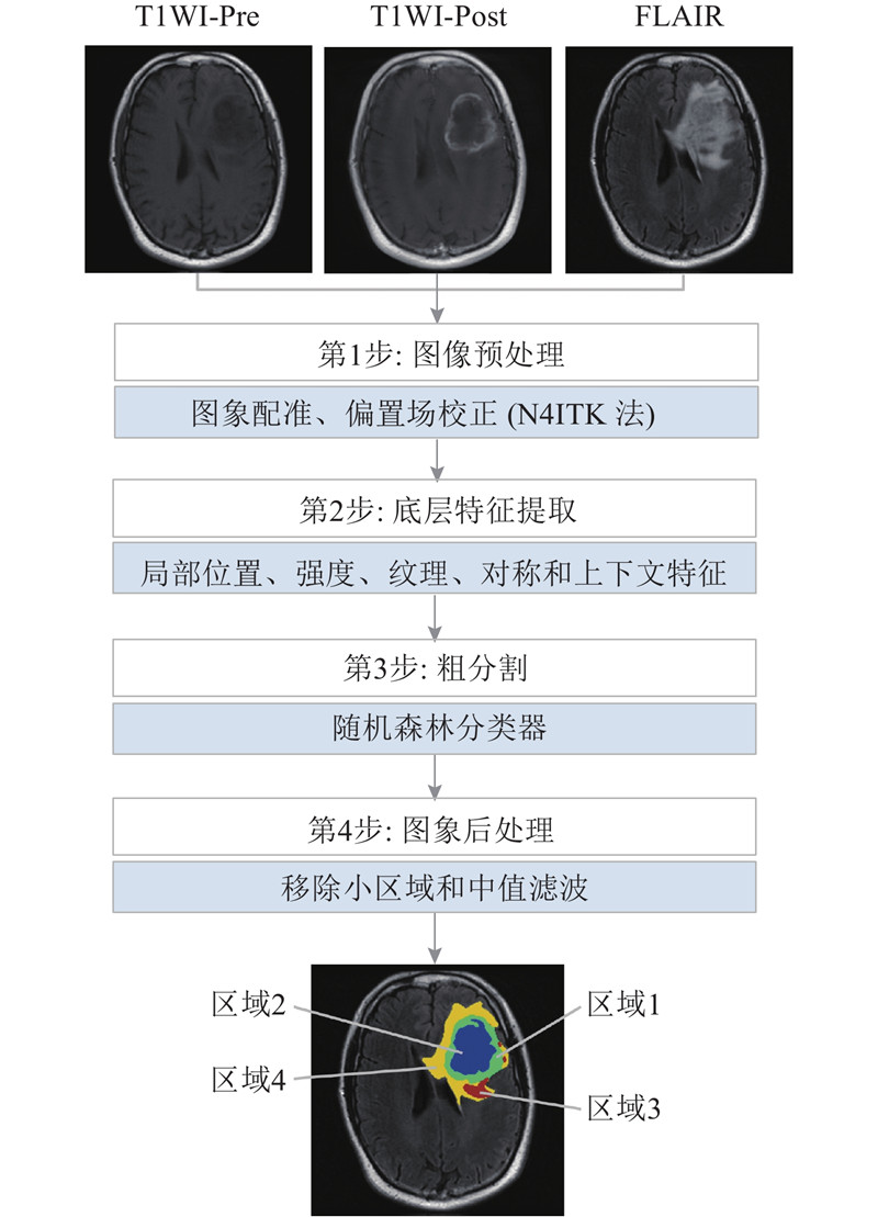
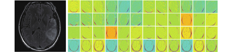
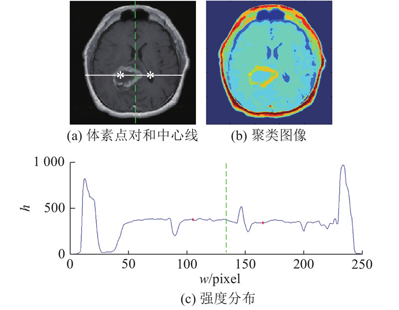
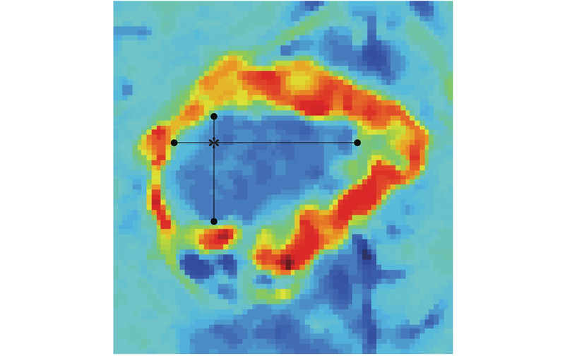
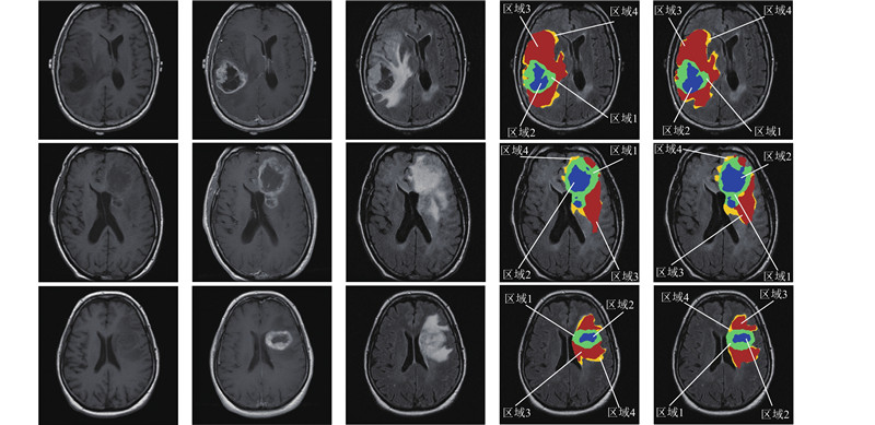
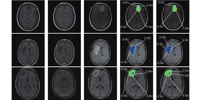
Abstract A glioblastoma multiforme (GBM) multi-modal magnetic resonance (MR) image automated segmentation algorithm based on hybrid features and prior knowledge was proposed, as most traditional GBM multi-modal MR image segmentation algorithms failed to subdivide the whole tumor into different sub-regions. The head region was adjusted to the approximate unrotated position once the GBM multi-modal MR image was registered, and the bias field correction was performed by the N4ITK method. A random forest classifier was applied to initially segment GBM multi-modal MR image after the extraction of the local location features, intensity features, texture features, symmetric features and contextual features of GBM multi-modal MR image. The final segmentation results were obtained by removing small regions and median filtering, based on the prior knowledge of the anatomical structure of GBM tumor. The Dice similarity coefficient was adopted as an evaluation metric, and the average Dice similarity coefficient values were 0.871 and 0.882 for segmenting the whole tumor in TCGA-GBM and CH-GBM databases by the proposed algorithm, respectively. Results indicated that the proposed method is suitable for clinical application of GBM multi-modal MR image segmentation task with relative high accuracy.
|
|
Received: 03 April 2018
Published: 21 February 2019
|
|
|
胶质母细胞瘤多模态磁共振图像自动分割
针对大多数传统胶质母细胞瘤(GBM)多模态磁共振(MR)图像分割算法未能将整个肿瘤细分为不同子区域的问题,提出基于混合特征和先验知识的GBM多模态MR图像自动分割算法. 配准GBM多模态MR图像,将头部区域方位调整到近似未旋转位置,并利用N4ITK法进行偏置场校正. 在提取GBM多模态MR图像局部位置特征、强度特征、纹理特征、对称特征和上下文特征后,应用随机森林分类器初步分割GBM多模态MR图像. 考虑GBM肿瘤解剖结构先验知识,移除小区域和中值滤波后得到最终分割结果. 以Dice相似性系数作为评价指标,利用所提出的算法对TCGA-GBM和CH-GBM数据库中整个肿瘤进行分割,获得的平均Dice相似性系数分别为0.871、0.882. 结果表明,该算法能以较高的准确率分割GBM多模态MR图像,适用于临床GBM多模态MR图像分割任务.
关键词:
胶质母细胞瘤(GBM),
多模态磁共振(MR)图像,
自动分割,
混合特征,
先验知识
|
|
| [1] |
PRASANNA P, PATEL J, PARTOVI S, et al Radiomic features from the peritumoral brain parenchyma on treatment-naive multi-parametric MR imaging predict long versus short-term survival in glioblastoma multiforme: preliminary findings[J]. European Radiology, 2017, 27 (10): 4198- 4199
doi: 10.1007/s00330-017-4815-y
|
|
|
| [2] |
RAVIKANTH R Advanced magnetic resonance imaging of glioblastoma multiforme[J]. Journal of Neurosciences in Rural Practice, 2017, 8 (3): 439- 440
doi: 10.4103/jnrp.jnrp_423_16
|
|
|
| [3] |
ZACHARAKI E, MORITA N, BHATT P, et al Survival analysis of patients with high-grade gliomas based on data mining of imaging variables[J]. American Journal of Neuroradiology, 2012, 33 (6): 1065- 1071
doi: 10.3174/ajnr.A2939
|
|
|
| [4] |
CLARK M C, HALL L O, GOLDGOF D B, et al Automatic tumor segmentation using knowledge-based techniques[J]. IEEE Transactions on Medical Imaging, 1998, 17 (2): 187- 201
doi: 10.1109/42.700731
|
|
|
| [5] |
FLETCHER-HEATH L M, HALL L O, GOLDGOF D B, et al Automatic segmentation of non-enhancing brain tumors in magnetic resonance images[J]. Artificial Intelligence in Medicine, 2001, 21 (1?3): 43- 63
doi: 10.1016/S0933-3657(00)00073-7
|
|
|
| [6] |
SHI J B, MALIK J Normalized cuts and image segmentation[J]. IEEE Transactions on Pattern Analysis and Machine Intelligence, 2000, 22 (8): 888- 905
doi: 10.1109/34.868688
|
|
|
| [7] |
CORSO J J, SHARON E, DUBE S, et al Efficient multilevel brain tumor segmentation with integrated bayesian model classification[J]. IEEE Transactions on Medical Imaging, 2008, 27 (5): 629- 640
doi: 10.1109/TMI.2007.912817
|
|
|
| [8] |
CHINNADURAI V, CHANDRASHEKHAR G D Neuro-levelset system based segmentation in dynamic susceptibility contrast enhanced and diffusion weighted magnetic resonance images[J]. Pattern Recognition, 2012, 45 (9): 3501- 3511
doi: 10.1016/j.patcog.2012.02.038
|
|
|
| [9] |
POPURI K, COBZAS D, MURTHA A, et al 3D variational brain tumor segmentation using Dirichlet priors on a clustered feature set[J]. International Journal of Computer Assisted Radiology and Surgery, 2012, 7 (4): 493- 506
doi: 10.1007/s11548-011-0649-2
|
|
|
| [10] |
POPE W B, SAYRE J, PERLINA A, et al MR imaging correlates of survival in patients with high-grade gliomas[J]. American Journal of Neuroradiology, 2005, 26 (10): 2466- 2474
|
|
|
| [11] |
GREVE D N, FISCHL B Accurate and robust brain image alignment using boundary-based registration[J]. Neuroimage, 2009, 48 (1): 63- 72
doi: 10.1016/j.neuroimage.2009.06.060
|
|
|
| [12] |
ALEXANDER V T, OLIVIER C, ISABELLE B Evaluation of the symmetry plane in 3D MR brain images[J]. Pattern Recognition Letters, 2003, 24 (14): 2219- 2233
doi: 10.1016/S0167-8655(03)00049-7
|
|
|
| [13] |
TUSTISON N J, AVANTS B B, COOK P A, et al N4ITK: improved N3 bias correction[J]. IEEE Transactions on Medical Imaging, 2010, 29 (6): 1310- 1320
doi: 10.1109/TMI.2010.2046908
|
|
|
| [14] |
VARMA M, ZISSERMAN A A statistical approach to texture classification from single images[J]. International Journal of Computer Vision, 2005, 62 (1): 61- 81
|
|
|
| [15] |
TUZIKOV A V, COLLIOT O, BLOCH I Evaluation of the symmetry plane in 3D MR brain images[J]. Pattern Recognition Letters, 2003, 24 (14): 2219- 2233
|
|
|
| [16] |
BAUER S, NOLTE L P, REYES M Fully automatic segmentation of brain tumor images using support vector machine classification in combination with hierarchical conditional random field regularization[J]. Medical Image Computing and Computer-Assisted Intervention, 2011, 14: 354- 361
|
|
|
| [17] |
GOOYA A, POHL K, BILELLO M, et al GLISTR: glioma image segmentation and registration[J]. IEEE Transactions on Medical Imaging, 2012, 31 (10): 1941- 1954
doi: 10.1109/TMI.2012.2210558
|
|
|
|
Viewed |
|
|
|
Full text
|
|
|
|
|
Abstract
|
|
|
|
|
Cited |
|
|
|
|
| |
Shared |
|
|
|
|
| |
Discussed |
|
|
|
|