|
|
|
| SA/PEO composite scaffold electrohydrodynamic direct writing fabrication and influencing factors analysis |
Lei SUN1( ),Chunjing WANG1,Wenlei ZHANG1,Zhipeng MA2,Yongqiang CHENG1 ),Chunjing WANG1,Wenlei ZHANG1,Zhipeng MA2,Yongqiang CHENG1 |
1. College of Integrated Circuits, Taiyuan University of Technology, Taiyuan 030024, China
2. School of Aeronautics and Astronautics, Zhejiang University, Hangzhou 310058, China |
|
|
|
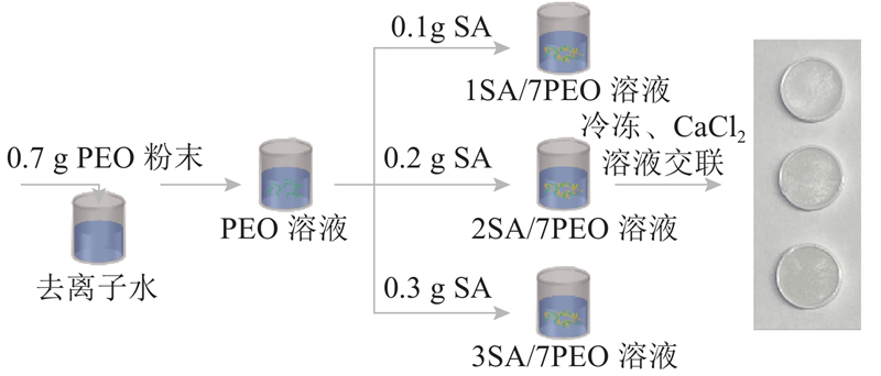
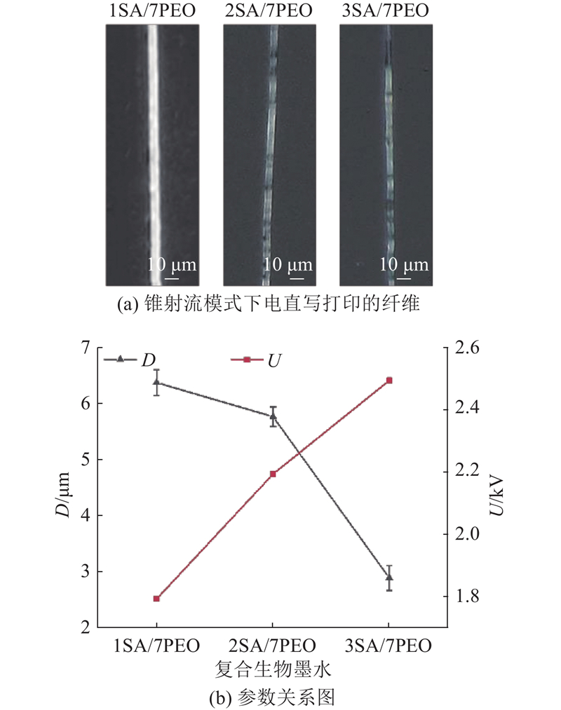
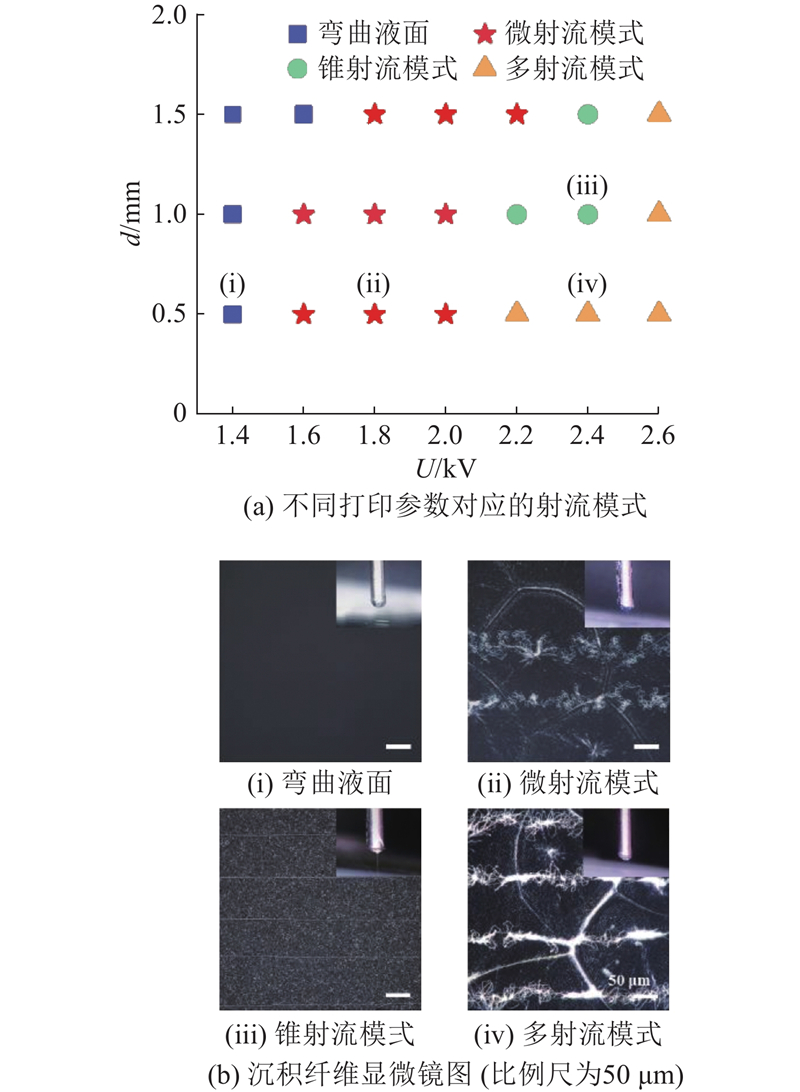
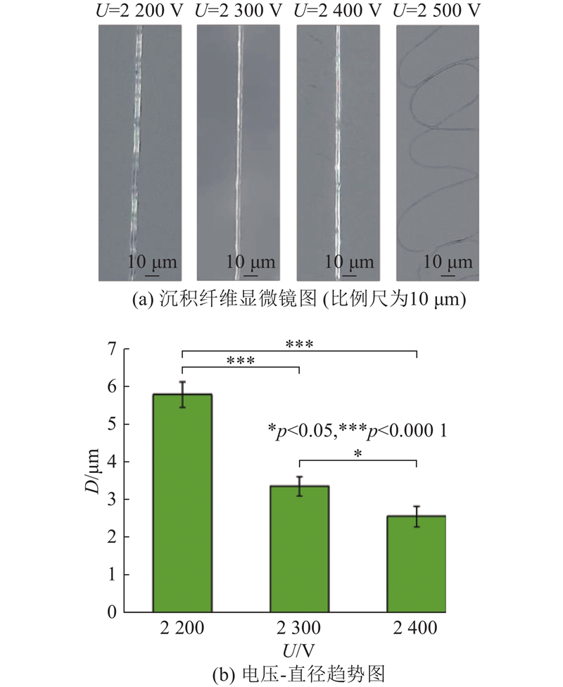
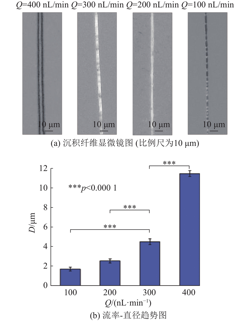
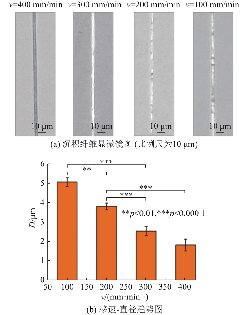
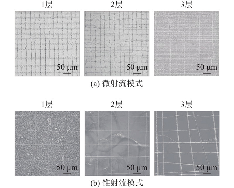
Abstract To improve the printing accuracy of electrohydrodynamic direct writing (EHDDW) in tissue engineering scaffold preparation, and to reduce equipment production costs, a low-cost EHDDW printing platform was constructed independently. Sodium alginate (SA)/polyethylene oxide (PEO) composite bioinks with varying concentration gradients were developed, and the biological characterization and printing test results reveal that the 2SA/7PEO composite bioink exhibits good cytocompatibility and EHDDW printing performance. Through theoretical analysis and experimental study, the influence mechanism of printing parameters on the fiber width of composite bioink was explored. Results show that increasing the voltage, decreasing the fluid flow rate, and accelerating the substrate movement speed can enhance the resolution of fibers. The SA/PEO composite bioink achieved a maximum printing resolution of 2.48±0.27 μm with an applied voltage of 2 400 V, a syringe pump flow rate of 200 nL/min, and a substrate movement speed of 300 mm/min. Single-layer/multi-layer grid micropatterns were prepared using fine-jet mode/cone-jet mode, proving that the self-constructed EHDDW printing platform has the potential to prepare complex and multilayered bio-microstructures.
|
|
Received: 14 June 2024
Published: 25 July 2025
|
|
|
| Fund: 山西省自然科学基金资助项目(202103021223069). |
海藻酸钠/聚氧化乙烯支架电直写成型及影响因素分析
为了提升电流体动力直写(EHDDW)在组织工程支架制备中的打印精度,降低设备及生产成本,自主搭建低成本电直写打印平台. 开发不同浓度梯度的海藻酸钠(SA)/聚氧化乙烯(PEO)复合生物墨水,生物学表征及打印测试结果表明,2SA/7PEO复合生物墨水具有良好的细胞相容性及电直写性能. 结合理论分析及实验探究打印参数对复合生物墨水沉积纤维宽度的影响机制,结果表明,增大电压、减小供液流率及提升基板移速均可提高纤维分辨率. 当电压为2 400 V,注射泵流率为200 nL/min,基板移速为300 mm/min时,SA/PEO复合生物墨水的最高打印精度为2.48±0.27 μm. 采用微射流模式/锥射流模式制备SA/PEO复合生物墨水单层/多层网格微图案,证明了自主搭建的电直写打印平台具备制备复杂、多层次生物微结构的潜力.
关键词:
电流体动力直写(EHDDW),
海藻酸钠(SA),
聚氧化乙烯(PEO),
锥射流模式,
组织工程
|
|
| [1] |
DAIKUARA L Y, CHEN X, YUE Z, et al 3D bioprinting constructs to facilitate skin regeneration[J]. Advanced Functional Materials, 2022, 32 (3): 2105080
doi: 10.1002/adfm.202105080
|
|
|
| [2] |
KANG M S, JANG J, JO H J, et al Advances and innovations of 3D bioprinting skin[J]. Biomolecules, 2023, 13 (1): 55
|
|
|
| [3] |
WU Y, CAI L, CHEN G, et al 3D printed, environment tolerant all-solid-state capacitive ionic skin[J]. Journal of Materials Chemistry A, 2022, 10 (35): 18218- 18225
doi: 10.1039/D2TA05388H
|
|
|
| [4] |
WANG S, LUO B, BAI B, et al 3D printed chondrogenic functionalized PGS bioactive scaffold for cartilage regeneration[J]. Advanced Healthcare Materials, 2023, 12 (27): 2301006
doi: 10.1002/adhm.202301006
|
|
|
| [5] |
MAIHEMUTI A, ZHANG H, LIN X, et al 3D-printed fish gelatin scaffolds for cartilage tissue engineering[J]. Bioactive Materials, 2023, 26: 77- 87
doi: 10.1016/j.bioactmat.2023.02.007
|
|
|
| [6] |
GOLD K A, SAHA B, RAJEEVA PANDIAN N K, et al 3D bioprinted multicellular vascular models[J]. Advanced Healthcare Materials, 2021, 10 (21): e2101141
doi: 10.1002/adhm.202101141
|
|
|
| [7] |
WANG P, SUN Y, SHI X, et al 3D printing of tissue engineering scaffolds: a focus on vascular regeneration[J]. Bio-Design and Manufacturing, 2021, 4 (2): 344- 378
doi: 10.1007/s42242-020-00109-0
|
|
|
| [8] |
SUN D, CHANG C, LI S, et al Near-field electrospinning[J]. Nano Letters, 2006, 6 (4): 839- 842
doi: 10.1021/nl0602701
|
|
|
| [9] |
HUANG Y, BU N, DUAN Y, et al Electrohydrodynamic direct-writing[J]. Nanoscale, 2013, 5 (24): 12007- 12017
doi: 10.1039/c3nr04329k
|
|
|
| [10] |
YIN Z, WANG D, GUO Y, et al Electrohydrodynamic printing for high resolution patterning of flexible electronics toward industrial applications[J]. InfoMat, 2024, 6 (2): e12505
doi: 10.1002/inf2.12505
|
|
|
| [11] |
MKHIZE N, MURUGAPPAN K, CASTELL M R, et al Electrohydrodynamic jet printed conducting polymer for enhanced chemiresistive gas sensors[J]. Journal of Materials Chemistry C, 2021, 9 (13): 4591- 4596
doi: 10.1039/D0TC05719C
|
|
|
| [12] |
COHEN T A, SHARP D, KLUHERZ K T, et al Direct patterning of perovskite nanocrystals on nanophotonic cavities with electrohydrodynamic inkjet printing[J]. Nano Letters, 2022, 22 (14): 5681- 5688
doi: 10.1021/acs.nanolett.2c00473
|
|
|
| [13] |
WU Y, FU C, QIAN S, et al Flexible and transparent W-band absorber fabricated by EHD printing technology[J]. IEEE Antennas and Wireless Propagation Letters, 2020, 19 (8): 1345- 1349
doi: 10.1109/LAWP.2020.3000786
|
|
|
| [14] |
DUAN Y, YANG W, XIAO J, et al High density, addressable electrohydrodynamic printhead made of a silicon plate and polymer nozzle structure[J]. Lab on a Chip, 2022, 22 (20): 3877- 3884
doi: 10.1039/D2LC00624C
|
|
|
| [15] |
YANG W, DUAN Y, GAO J, et al Crosstalk elimination for large-scale, high-density electrohydrodynamic printing via optimization of nozzle material and structure[J]. Additive Manufacturing, 2023, 77: 103815
doi: 10.1016/j.addma.2023.103815
|
|
|
| [16] |
HE J, HAO G, MENG Z, et al Expanding melt-based electrohydrodynamic printing of highly-ordered microfibrous architectures to cm-height via in situ charge neutralization[J]. Advanced Materials Technologies, 2022, 7 (7): 2101197
doi: 10.1002/admt.202101197
|
|
|
| [17] |
LI K, WANG D, ZHAO K, et al Electrohydrodynamic jet 3D printing of PCL/PVP composite scaffold for cell culture[J]. Talanta, 2020, 211: 120750
doi: 10.1016/j.talanta.2020.120750
|
|
|
| [18] |
LI K, WANG D, ZHANG F, et al Tip-viscid electrohydrodynamic jet 3D printing of composite osteochondral scaffold[J]. Nanomaterials, 2021, 11 (10): 2694
doi: 10.3390/nano11102694
|
|
|
| [19] |
YAO C, QIU Z, LI X, et al Electrohydrodynamic printing of microfibrous architectures with cell-scale spacing for improved cellular migration and neurite outgrowth[J]. Small, 2023, 19 (19): 2207331
doi: 10.1002/smll.202207331
|
|
|
| [20] |
QIU Z, ZHU H, WANG Y, et al Functionalized alginate-based bioinks for microscale electrohydrodynamic bioprinting of living tissue constructs with improved cellular spreading and alignment[J]. Bio-Design and Manufacturing, 2023, 6 (2): 136- 149
doi: 10.1007/s42242-022-00225-z
|
|
|
| [21] |
YEO M, KIM G Electrohydrodynamic-direct-printed cell-laden microfibrous structure using alginate-based bioink for effective myotube formation[J]. Carbohydrate Polymers, 2021, 272: 118444
doi: 10.1016/j.carbpol.2021.118444
|
|
|
| [22] |
FANG Y, WANG C, LIU Z, et al 3D printed conductive multiscale nerve guidance conduit with hierarchical fibers for peripheral nerve regeneration[J]. Advanced Science, 2023, 10 (12): 2205744
doi: 10.1002/advs.202205744
|
|
|
| [23] |
ZHANG Z, JØRGENSEN M L, WANG Z, et al 3D anisotropic photocatalytic architectures as bioactive nerve guidance conduits for peripheral neural regeneration[J]. Biomaterials, 2020, 253: 120108
doi: 10.1016/j.biomaterials.2020.120108
|
|
|
| [24] |
WANG J, WANG H, MO X, et al Reduced graphene oxide-encapsulated microfiber patterns enable controllable formation of neuronal-like networks[J]. Advanced Materials, 2020, 32 (40): 2004555
doi: 10.1002/adma.202004555
|
|
|
| [25] |
FUH Y K, WU Y C, HE Z Y, et al The control of cell orientation using biodegradable alginate fibers fabricated by near-field electrospinning[J]. Materials Science and Engineering: C, 2016, 62: 879- 887
doi: 10.1016/j.msec.2016.02.028
|
|
|
| [26] |
郑高峰, 钟炜政, 姜佳昕, 等. 基于PEO电纺膜基底的近场直写聚焦及微图案脱离方法: 201910078347.0 [P]. 2020–02–10.
|
|
|
| [27] |
ANDRUKHOV O, HUBER R, SHI B, et al Proliferation, behavior, and differentiation of osteoblasts on surfaces of different microroughness[J]. Dental Materials, 2016, 32 (11): 1374- 1384
doi: 10.1016/j.dental.2016.08.217
|
|
|
| [28] |
XIE C, GAO Q, WANG P, et al Structure-induced cell growth by 3D printing of heterogeneous scaffolds with ultrafine fibers[J]. Materials and Design, 2019, 181: 108092
doi: 10.1016/j.matdes.2019.108092
|
|
|
|
Viewed |
|
|
|
Full text
|
|
|
|
|
Abstract
|
|
|
|
|
Cited |
|
|
|
|
| |
Shared |
|
|
|
|
| |
Discussed |
|
|
|
|