| Computer Science and Artificial Intelligence |
|


|
|
|
| Classification of renal tumor histology subtypes using radiomics |
Yi YANG1,2( ),Xu-sheng QIAN2,Zhi-yong ZHOU2,Jian-bing ZHU3,4,Jun-kang SHEN5,Ya-kang DAI2,*() ),Xu-sheng QIAN2,Zhi-yong ZHOU2,Jian-bing ZHU3,4,Jun-kang SHEN5,Ya-kang DAI2,*() |
1. University of Science and Technology of China, Hefei 230000, China
2. Suzhou Institute of Biomedical Engineering and Technology, Chinese Academy of Sciences, Suzhou 215163, China
3. Suzhou Science and Technology Town Hospital, Suzhou 215163, China
4. Suzhou Science and Technology Hospital Affiliated to Nanjing Medical University, Suzhou 215163, China
5. Second Affiliated Hospital of Suzhou University, Suzhou 215163, China |
|
|
|
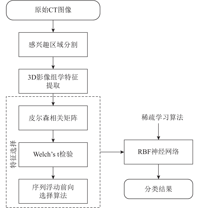
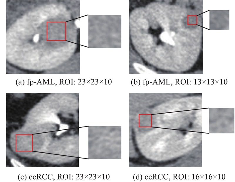
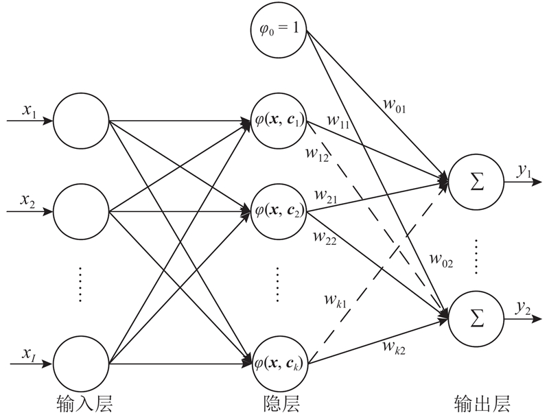
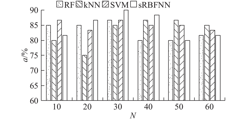
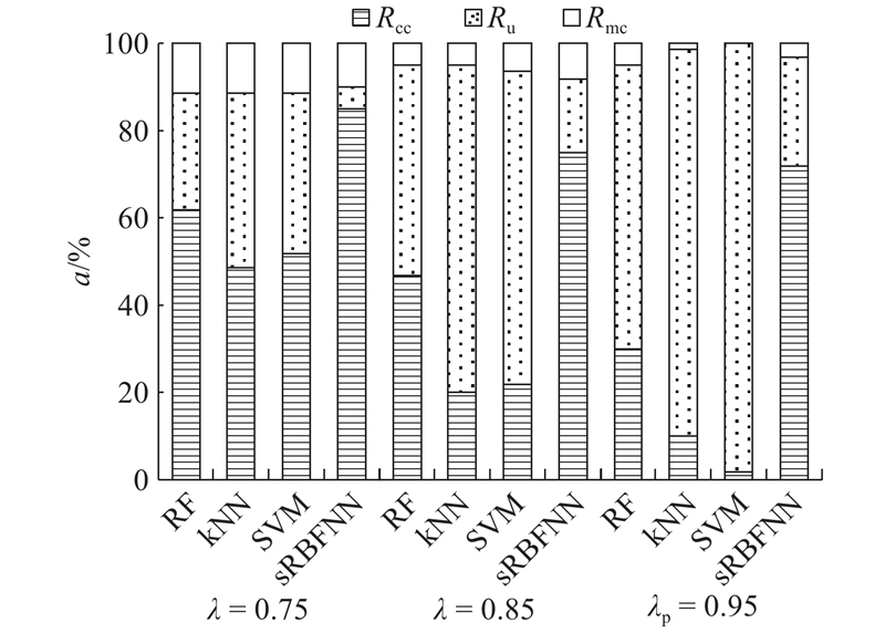
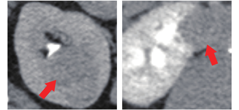
Abstract A CT-based radiomics model was proposed to increase the accuracy of preoperative noninvasive differentiation of fp-AML from ccRCC. There were 774 three-dimensional radiomics features extracted from CT images. The feature selection was carried by three steps: Pearson’s correlation matrices were calculated to remove redundant features; Welch’s t-test was used to determine the statistically significant features; and sequential forward floating selection method was utilized to select the discriminative features. The sparse radial basis function neural network was employed for classification. Results show that the radiomics model yields accuracy, sensitivity, specificity, and the area under the receiver operating characteristic curves of 90.00%, 66.67%, 100.0%, and 0.9173, respectively. The reliability of model was assessed by probabilistic outputs of classifiers. When the probability threshold is 0.95, the model obtains confident classification accuracy, undecided rate, and misclassification rate of 71.67%, 25.00%, and 3.33%, respectively. Results demonstrate that the proposed radiomics model can achieve reliable discrimination of fp-AML from ccRCC.
|
|
Received: 22 February 2019
Published: 17 December 2019
|
|
|
|
Corresponding Authors:
Ya-kang DAI
E-mail: yangyi129@icloud.com;daiyk@sibet.ac.cn
|
采用影像组学的肾肿瘤组织学亚型分类
为了在术前更准确、非侵入地鉴别乏脂肪血管平滑肌脂肪瘤(fp-AML)和肾透明细胞癌(ccRCC),提出一种基于CT图像的影像组学模型. 从CT图像中提取774个三维的影像组学特征;分三步进行特征选择:计算皮尔森相关矩阵剔除冗余特征,使用Welch’s t检验确定具有显著差异的特征,利用序列浮动前向选择算法选择具有鉴别能力的特征;使用基于稀疏学习的径向基函数神经网络进行分类. 结果表明:该模型获得的正确率、敏感度、特异性和受试者工作特征曲线下面积分别为90.00%、66.67%、100.0%和0.9173. 利用分类器的输出概率进行模型的可靠性评估,当概率阈值为0.95时,该模型获得的自信正确率、未定率和错分率分别为71.67%、25.00%和3.33%,结果表明所提出的影像组学模型能可靠地对fp-AML和ccRCC进行分类.
关键词:
计算机辅助诊断,
乏脂肪血管平滑肌脂肪瘤,
肾透明细胞癌,
影像组学,
径向基函数神经网络
|
|
| [1] |
HSIEH J J, PURDUE P M, SIGNORETTI S, et al Renal cell carcinoma[J]. Nature Reviews Disease Primers, 2017, 3: 17009
doi: 10.1038/nrdp.2017.9
|
|
|
| [2] |
WORLD Cancer Research Fund International. Kidney cancer statistics [J/OL]. Available at https://www.wcrf.org/dietandcancer/cancer-trends/kidney-cancer-statistics, January 2018.
|
|
|
| [3] |
WEI J, ZHAO J, ZHANG X, et al Analysis of dual energy spectral CT and pathological grading of clear cell renal cell carcinoma (ccRCC)[J]. PLOS ONE, 2018, 13 (5): e0195699
doi: 10.1371/journal.pone.0195699
|
|
|
| [4] |
JINZAKI M, SIVERMAN S G, and TANIMOTO A Angiomyolipoma that do not contain fat attenuation at unenhanced CT[J]. Radiology, 2005, 234 (1): 311
doi: 10.1148/radiol.2341041128
|
|
|
| [5] |
PRASAD S R, SURABHI V R, MENIAS C O, et al Benign renal neoplasms in adults: cross-sectional imaging findings[J]. American Journal of Roentgenology, 2008, 190 (1): 158- 164
doi: 10.2214/AJR.07.2724
|
|
|
| [6] |
HALPENNY D, SNOW A, MCNEILL G, et al The radiological diagnosis and treatment of renal angiomyolipoma-current status[J]. Clinical Radiology, 2010, 65 (2): 99- 108
doi: 10.1016/j.crad.2009.09.014
|
|
|
| [7] |
LANE B R, AYDIN H, DANFORTH T L, et al Clinical correlates of renal angiomyolipoma subtypes in 209 patients: classic, fat poor, tuberous sclerosis associated and epithelioid[J]. The Journal of Urology, 2008, 180 (3): 836- 843
doi: 10.1016/j.juro.2008.05.041
|
|
|
| [8] |
FENG Z, RONG P, CAO P, et al Machine learning-based quantitative texture analysis of CT images of small renal masses: differentiation of angiomyolipoma without visible fat from renal cell carcinoma[J]. European Radiology, 2018, 28 (4): 1625- 1633
doi: 10.1007/s00330-017-5118-z
|
|
|
| [9] |
LEE H S, HONG H, JUNG D C, et al Differentiation of fat-poor angiomyolipoma from clear cell renal cell carcinoma in contrast-enhanced MDCT images using quantitative feature classification[J]. Medical Physics, 2017, 44 (7): 3604- 3614
doi: 10.1002/mp.2017.44.issue-7
|
|
|
| [10] |
MOLINA D, JULIáN P, BELéN L, et al Tumour heterogeneity in glioblastoma assessed by MRI texture analysis: a potential marker of survival[J]. The British Journal of Radiology, 2016, 89 (1064): 20160242
doi: 10.1259/bjr.20160242
|
|
|
| [11] |
LAMBIN P, LEIJENAAR R T H, DEIST T M, et al Radiomics: the bridge between medical imaging and personalized medicine[J]. Nature Reviews Clinical Oncology, 2017, 14: 749
doi: 10.1038/nrclinonc.2017.141
|
|
|
| [12] |
ELISABETTA D B, BUDA A, GUERRA L, et al Radiomics of the primary tumour as a tool to improve 18F-FDG-PET sensitivity in detecting nodal metastases in endometrial cancer[J]. EJNMMI Research, 2018, 8 (1): 86
doi: 10.1186/s13550-018-0441-1
|
|
|
| [13] |
FERREIRA JUNIOR J R, KOENIGKAM-SANTOS M, CIPRIANO F E G, et al Radiomics-based features for pattern recognition of lung cancer histopathology and metastases[J]. Computer Methods and Programs in Biomedicine, 2018, 159: 23- 30
doi: 10.1016/j.cmpb.2018.02.015
|
|
|
| [14] |
SAPATE S G, MAHAJAN A, TALBAR S N, et al Radiomics based detection and characterization of suspicious lesions on full field digital mammograms[J]. Computer Methods and Programs in Biomedicine, 2018, 163: 1- 20
doi: 10.1016/j.cmpb.2018.05.017
|
|
|
| [15] |
MENG X, XIA W, XIE P, et al Preoperative radiomic signature based on multiparametric magnetic resonance imaging for noninvasive evaluation of biological characteristics in rectal cancer[J]. European Radiology, 2018, 1- 10
|
|
|
| [16] |
QIAN X, HUANG H, CHEN X, et al Efficient construction of sparse radial basis function neural networks using L1-regularization[J]. Neural Networks, 2017, 94: 239- 254
doi: 10.1016/j.neunet.2017.07.004
|
|
|
| [17] |
LUBNER M G, SMITH A D, SANDRASEGARAN K, et al CT texture analysis: definitions, applications, biologic correlates, and challenges[J]. RadioGraphics, 2017, 37 (5): 1483- 1503
doi: 10.1148/rg.2017170056
|
|
|
| [18] |
ZWANENBURG A, LEGER S, VALLIèRES M, et al. Image biomarker standardisation initiative-feature definitions [J/OL]. arXiv eprints, 2016. https://ui.adsabs.harvard.edu/abs/2016arXiv161207003Z
|
|
|
| [19] |
AERTS H J, VELAZQUEZ E R, LEIJENAAR R T, et al Decoding tumour phenotype by noninvasive imaging using a quantitative radiomics approach[J]. Nature Communications, 2014, 5: 4006
doi: 10.1038/ncomms5006
|
|
|
| [20] |
HARIKUMAR R and VINOTH KUMAR B Performance analysis of neural networks for classification of medical images with wavelets as a feature extractor[J]. International Journal of Imaging Systems and Technology, 2015, 25 (1): 33- 40
doi: 10.1002/ima.v25.1
|
|
|
| [21] |
PEARSON K Note on regression and inheritance in the case of two parents[J]. Proceedings of the Royal Society of London, 1895, 58: 240- 242
doi: 10.1098/rspl.1895.0041
|
|
|
| [22] |
DERRICK B and WHITE P Why Welch’s test is type I error robust[J]. The Quantitative Methods in Psychology, 2016, 12 (1): 30- 38
doi: 10.20982/tqmp.12.1.p030
|
|
|
| [23] |
AHA D W, BANKERT R L. A Comparative Evaluation of Sequential Feature Selection Algorithms [M]. New York: Springer, 1996: 199-206.
|
|
|
| [24] |
FALLAHPOUR S, LAKVAN E N, and ZADEH M H Using an ensemble classifier based on sequential floating forward selection for financial distress prediction problem[J]. Journal of Retailing and Consumer Services, 2017, 34: 159- 167
doi: 10.1016/j.jretconser.2016.10.002
|
|
|
| [25] |
BUGDOL M D, BUGDOL M N, LIPOWICZ A M, et al Prediction of menarcheal status of girls using voice features[J]. Computers in Biology and Medicine, 2018, 100: 296- 304
doi: 10.1016/j.compbiomed.2017.11.005
|
|
|
| [26] |
PARMAR C, GROSSMANN P, BUSSINK J, et al Machine learning methods for quantitative radiomic biomarkers[J]. Scientific Reports, 2015, 5: 13087
doi: 10.1038/srep13087
|
|
|
| [27] |
JOSHI A, METHA A Analysis of K-nearest neighbor technique for breast cancer disease classification[J]. International Journal of Recent Scientific Research, 2018, 9 (4): 26126- 26130
|
|
|
| [28] |
JOSé LUIS R, MANEL M, JORDI M, et al. Support vector machine and kernel classification algorithms [C] // Digital Signal Processing with Kernel Methods. New Jersey: Wiley-IEEE Press, 2018: 672.
|
|
|
| [29] |
CHENG J Z, NI D, CHOU Y H, et al Computer-aided diagnosis with deep learning architecture: applications to breast lesions in US images and pulmonary nodules in CT scans[J]. Scientific Reports, 2016, 6: 24454
doi: 10.1038/srep24454
|
|
|
| [30] |
LEE H, HONG H, KIM J, et al Deep feature classification of angiomyolipoma without visible fat and renal cell carcinoma in abdominal contrast-enhanced CT images with texture image patches and hand-crafted feature concatenation[J]. Medical Physics, 2018, 45 (4): 1550
doi: 10.1002/mp.2018.45.issue-4
|
|
|
| [31] |
CHAPELLE O, VAPNIK V, BOUSQUET O, et al Choosing multiple parameters for support vector machines[J]. Machine Learning, 2002, 46 (1): 131- 159
|
|
|
| [32] |
LUKAS L, DEVOS A, SUYKENS J A K, et al Brain tumor classification based on long echo proton MRS signals[J]. Artificial Intelligence in Medicine, 2004, 31 (1): 73- 89
doi: 10.1016/j.artmed.2004.01.001
|
|
|
|
Viewed |
|
|
|
Full text
|
|
|
|
|
Abstract
|
|
|
|
|
Cited |
|
|
|
|
| |
Shared |
|
|
|
|
| |
Discussed |
|
|
|
|