| 计算机技术、自动化技术 |
|

|
|
|
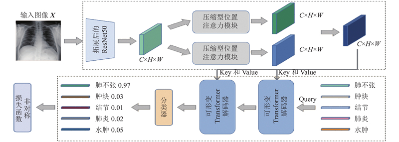
| 可形变Transformer辅助的胸部X光影像疾病诊断模型 |
胡锦波1( ),聂为之1,宋丹1,*(),高卓2,白云鹏3,赵丰3 ),聂为之1,宋丹1,*(),高卓2,白云鹏3,赵丰3 |
1. 天津大学 电气自动化与信息工程学院,天津 300072
2. 长春职业技术学院 信息学院,吉林 长春 130033
3. 天津市胸科医院 心血管外科,天津 300222 |
|
| Chest X-ray imaging disease diagnosis model assisted by deformable Transformer |
| Jin-bo HU1(),Wei-zhi NIE1,Dan SONG1,*(),Zhuo GAO2,Yun-peng BAI3,Feng ZHAO3 |
1. School of Electrical and Information Engineering, Tianjin University, Tianjin 300072, China
2. School of Information, Changchun Polytechnic, Changchun 130033, China
3. Department of Cardiovascular Surgery, Tianjin Chest Hospital, Tianjin 300222, China |
引用本文:
胡锦波,聂为之,宋丹,高卓,白云鹏,赵丰. 可形变Transformer辅助的胸部X光影像疾病诊断模型[J]. 浙江大学学报(工学版), 2023, 57(10): 1923-1932.
Jin-bo HU,Wei-zhi NIE,Dan SONG,Zhuo GAO,Yun-peng BAI,Feng ZHAO. Chest X-ray imaging disease diagnosis model assisted by deformable Transformer. Journal of ZheJiang University (Engineering Science), 2023, 57(10): 1923-1932.
链接本文:
https://www.zjujournals.com/eng/CN/10.3785/j.issn.1008-973X.2023.10.002
或
https://www.zjujournals.com/eng/CN/Y2023/V57/I10/1923
|
| 1 |
石连红 放射科医生的透视眼—CT与核磁共振[J]. 特别健康, 2020, 8: 35
SHI Lian-hong The fluoroscopy eye of the radiologist -CI and MRI[J]. Special Health, 2020, 8: 35
|
| 2 |
ELDAHSHAN E S A, MOHSEN H M, REVETT K, et al Computer-aided diagnosis of human brain tumor through MRI: a survey and a new algorithm[J]. Expert Systems with Applications, 2014, 41 (11): 5526- 5545
doi: 10.1016/j.eswa.2014.01.021
|
| 3 |
CHEN J, YU H, FENG R, et al. Flow-Mixup: classifying multi-labeled medical images with corrupted labels [C]// BIBM. Piscataway: IEEE, 2020: 534-541.
|
| 4 |
于玉海, 林鸿飞, 孟佳娜, 等 跨模态多标签生物医学图像分类建模识别[J]. 中国图像图形学报, 2018, 23 (6): 917- 927
YU Yu-hai, LIN Hong-fei, MENG Jia-na, et al Cross- modal multi-label biomedical image classification modeling and recognition[J]. Journal of Image and Graphics, 2018, 23 (6): 917- 927
|
| 5 |
LUO Y, TAO D, GENG B, et al Manifold regularized multitask learning for semi-supervised multilabel image classification[J]. IEEE Transactions on Image Processing, 2013, 22 (2): 523- 536
doi: 10.1109/TIP.2012.2218825
|
| 6 |
WEI YUNCHAO, XIA WEI, LIN MIN, et al HCP: a flexible CNN framework for multi-label image classification[J]. IEEE Transactions on Pattern Analysis and Machine Intelligence, 2016, 38 (9): 1901- 1907
doi: 10.1109/TPAMI.2015.2491929
|
| 7 |
BENBARUCH E, RIDNIK T, ZAMIR N, et al. Asymmetric loss for multi-label classification [C]// ICCV. Piscataway: IEEE, 2021: 82- 91.
|
| 8 |
GUO G R, ARNALDO M, ELIE B A, et al Artificial intelligence in healthcare: review and prediction case studies[J]. Engineering, 2020, 6 (3): 291- 301
doi: 10.1016/j.eng.2019.08.015
|
| 9 |
MILLER D D, BROWN E W Artificial intelligence in medical practice: the question to the answer[J]. The American Journal of Medicine, 2018, 131 (2): 129- 133
doi: 10.1016/j.amjmed.2017.10.035
|
| 10 |
ESTEVA A, KUPREL B, NOVOA R A Dermatologist level classification of skin cancer with deep neural networks[J]. Oncologie, 2017, 19 (11/12): 407- 408
doi: 10.1007/s10269-017-2730-4
|
| 11 |
MCKINNEY S M, SIENIEK M, GODBOLE V, et al International evaluation of an AI system for breast cancer screening[J]. Nature, 2020, 577 (7788): 89- 94
doi: 10.1038/s41586-019-1799-6
|
| 12 |
ZHANG J W, HE J T, CHEN T F, et al Abnormal region detection in cervical smear images based on fully convolutional network[J]. IET Image Processing, 2019, 13 (4): 583- 590
doi: 10.1049/iet-ipr.2018.6032
|
| 13 |
WANG X S, PENG Y F, LU L, et al. ChestX-ray8: hospital-scale chest X-ray database and benchmarks on weakly-supervised classification and localization of common thorax diseases [C]// 30th IEEE/CVF Conference on Computer Vision and Pattern Recognition. Piscataway: IEEE, 2017: 3462-3471.
|
| 14 |
HE K M, ZHANG X Y, REN S Q. Deep residual learning for image recognition [C]// IEEE Conference on Computer Vision and Pattern Recognition. Piscataway: IEEE, 2016: 770-778.
|
| 15 |
LI Y, ERIC P, DMITRY D, et al. Learning to diagnose from scratch by exploiting dependencies among labels [C]// IEEE Conference on Computer Vision and Pattern Recognition. Piscataway: IEEE, 2017: 7925-7937.
|
| 16 |
HUANG G, LIU Z, LAURENS V D M, et al. Densely connected convolutional networks [C]// IEEE Conference on Computer Vision and Pattern Recognition. Piscataway: IEEE, 2017: 2261-2269.
|
| 17 |
GUENDEL S, GRBIC S, GEORGESCU B, et al. Learning to recognize abnormalities in chest X-rays with location-aware Dense Networks [C]// Iberoamerican Congress on Pattern Recognition. Berlin: Springer, 2018: 757-765.
|
| 18 |
CHEN X L, GUPTA ABHINAV. Webly supervised learning of convolutional networks [C]// IEEE International Conference on Computer Vision. Piscataway: IEEE, 2016: 1431- 1439.
|
| 19 |
RAJPURKAR P, IRVIN J, ZHU K, et al. CheXNet: radiologist-level pneumonia detection on chest X-Rays with deep learning [C]// IEEE Conference on Computer Vision and Pattern Recognition. Piscataway: IEEE, 2017: 2698-2705.
|
| 20 |
LIU S L, ZHANG L, YANG X, et al. Query2Label: a simple transformer way to multi-label classification [C]// IEEE Conference on Computer Vision and Pattern Recognition. Piscataway: IEEE, 2021: 391-407.
|
| 21 |
BELLO I, FEDUS W, DU X, et al Revisiting ResNets: improved training and scaling strategies[J]. Advances in Neural Information Processing Systems, 2021, 34: 22614- 22627
|
| 22 |
LIN T Y, GOYAL P, GIRSHICK R, et al Focal loss for dense object detection[J]. IEEE Transactions on Pattern Analysis and Machine Intelligence, 2020, 42 (2): 318- 327
doi: 10.1109/TPAMI.2018.2858826
|
| 23 |
IRVIN J, RAJPURKAR P, KO M, et al. CheXpert: a large chest radiograph dataset with uncertainty labels and expert comparison [C]// 33rd AAAI Conference on Artificial Intelligence. Menlo Park: AAAI, 2019: 590-597.
|
| 24 |
YAN C, YAO J, LI R, et al. Weakly supervised deep learning for thoracic disease classification and localization on chest X-Rays [C]// Proc of ICBCB. New York: ACM, 2018: 103-110.
|
| 25 |
MA C, WANG H, HOI S C H. Multi-label thoracic disease image classification with cross-attention networks. international conference on medical image computing and computer-assisted intervention [C]// International Conference on Medical Image Computing and Computer Assisted Intervention. Berlin: Springer, 2019: 730-738.
|
| 26 |
TEIXEIRA V, BRAZ L, PEDRINI H, et al. DuaLAnet: dual lesion attention network for thoracic disease classification in chest X-rays [C]// International Conference on Systems, Signals and Image Processing. Piscataway: IEEE, 2020: 69-74.
|
| 27 |
LUO L, YU L, CHEN H, et al Deep mining external imperfect data for chest x-ray disease screening[J]. IEEE Trans Med Image, 2020, 39 (11): 3583- 3594
doi: 10.1109/TMI.2020.3000949
|
| 28 |
GUAN Q, HUANG Y, LUO Y, et al Discriminative feature learning for thorax disease classification in chest X-ray images[J]. IEEE Transactions on Image Processing, 2021, 99: 1- 2
|
| 29 |
PHAM H H, LE T T, TRAN D Q, et al Interpreting chest x-rays via cnns that exploit hierarchical disease dependencies and uncertainty labels[J]. Neurocomputing, 2021, 437: 186- 194
doi: 10.1016/j.neucom.2020.03.127
|
| 30 |
KAMAL U, ZUNAED M, NIZAM N B, et al Anatomy X-Net: a semi-supervised anatomy aware convolutional neural network for thoracic disease classification[J]. IEEE Journal of Biomedical and Health Informatics, 2021, 26 (11): 5518- 5528
|
|
Viewed |
|
|
|
Full text
|
|
|
|
|
Abstract
|
|
|
|
|
Cited |
|
|
|
|
| |
Shared |
|
|
|
|
| |
Discussed |
|
|
|
|