|
|
|
| Biological 3D printer and topography detection of printing model |
Da-peng BAI1( ),Bin ZHANG1,*(),Hao-cen HONG1,Yang LI1,Qing-hua JI2,Hua-yong YANG1 ),Bin ZHANG1,*(),Hao-cen HONG1,Yang LI1,Qing-hua JI2,Hua-yong YANG1 |
1. State Key Laboratory of Fluid Power and Mechatronic Systems, School of Mechanical Engineering, Zhejiang University, Hangzhou 310027, China
2. Binhai Industrial Technology Research Institute of Zhejiang University, Tianjin 300450, China |
|
|
|
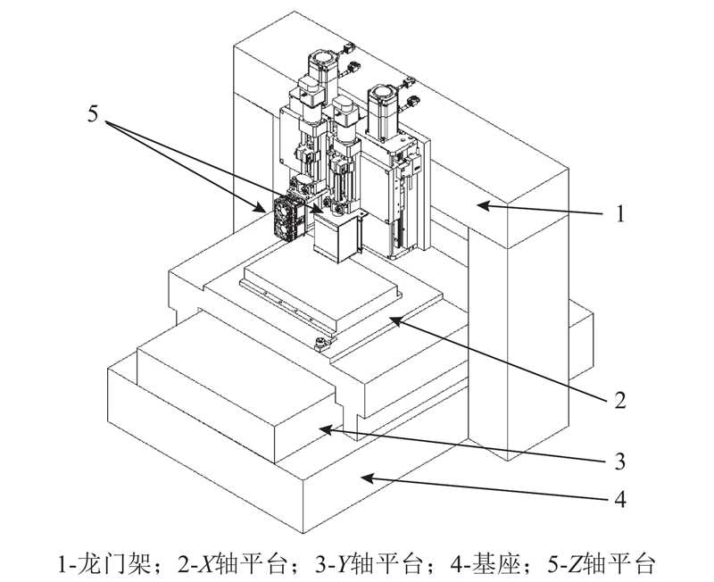
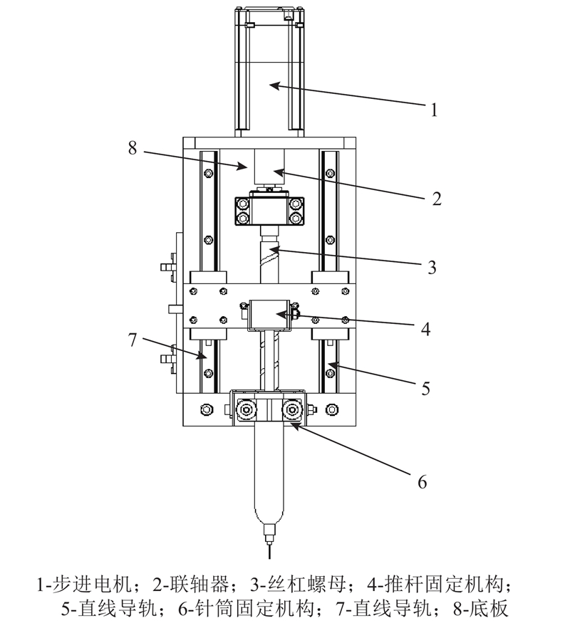
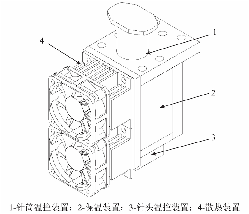
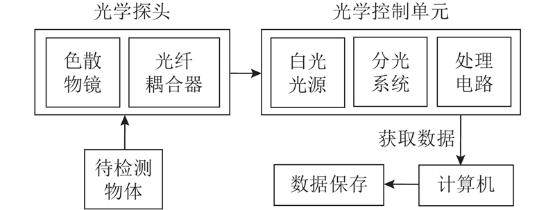
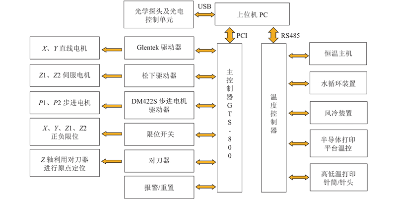
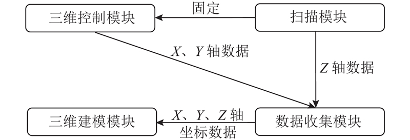
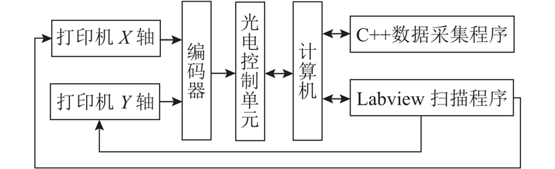
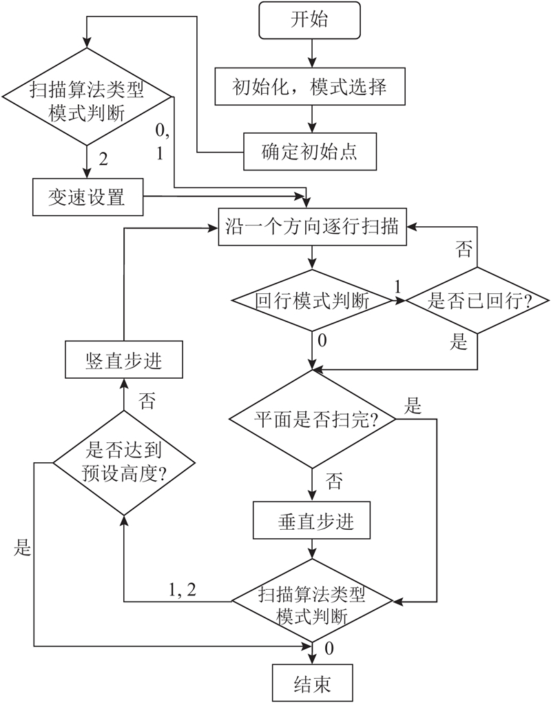
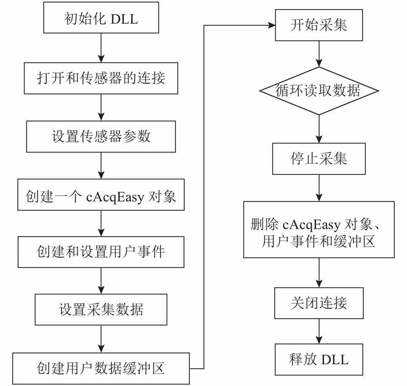
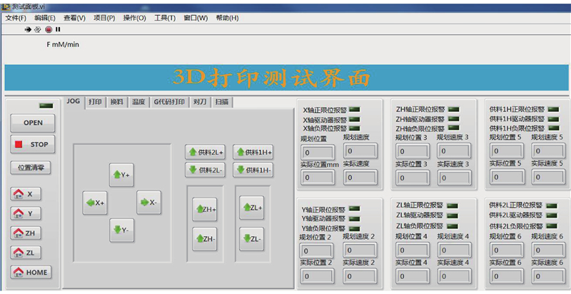
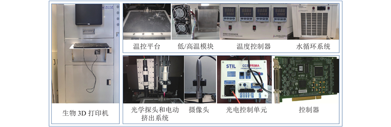
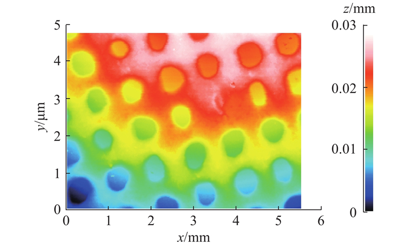
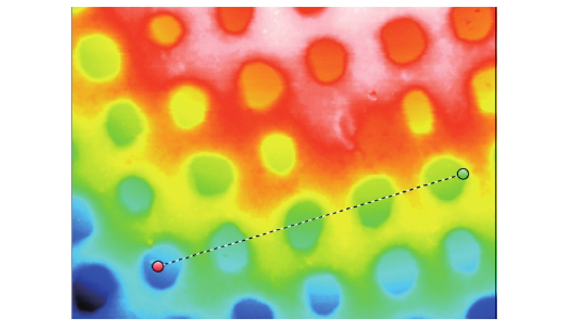
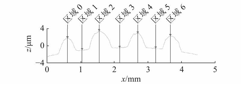
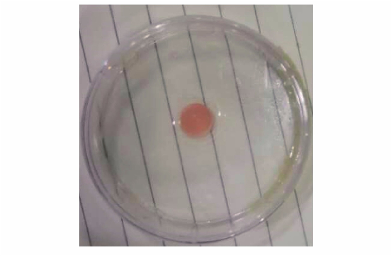
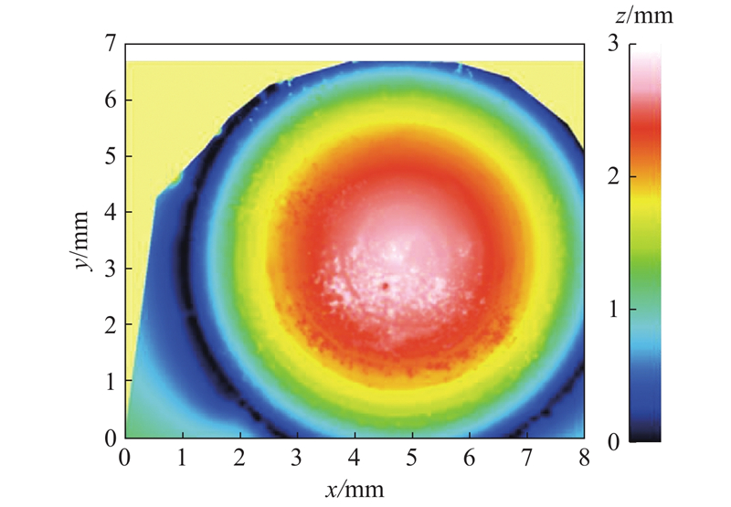
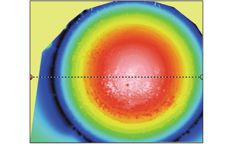
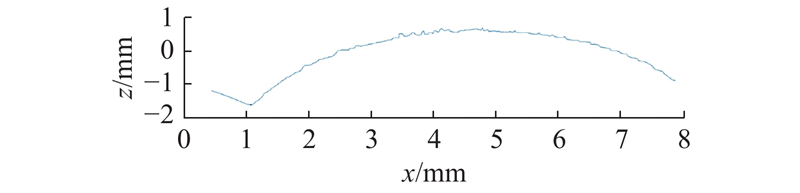
Abstract An integrated biological 3D printer inspection system was designed in order to observe the printing process of 3D biological printer in real time as well as detect and reconstruct the surface topography of printing model. The online inspection of printing model was realized based on the printing needles with video monitoring. The dispersion confocal displacement measurement technology was adopted to measure the position information of X-axis and Y-axis and the height information of Z-axis. The topography reconstruction work of printing model was processed by MountainsMap. The upper computer operating system was developed by LabVIEW, in which the printing model was mapped with G-code to visualize printing track to ensure the accuracy of the printing model when the program runs to the current code segment. The online real-time detecting function of the proposed biological 3D printer was verified by experiments, and the surface topography detection of grid shaped and convex shaped models was conducted respectively. Results show that the surface topography information of printing model can be reconstructed by the detection system. The visualization data of the surface topography features of the printing model provides data support for the construction of accurate printing models, and the computer vision technology provides an effective detection method for high-precision biological 3D printing.
|
|
Received: 08 August 2020
Published: 09 March 2021
|
|
|
| Fund: 国家重点研发计划资助项目(2018YFA0703000) |
|
Corresponding Authors:
Bin ZHANG
E-mail: bdp.2008@163.com;zbzju@163.com
|
生物3D打印装置及打印模型形貌检测
为了能够实时观测生物3D打印全过程,并对打印模型的形貌进行检测及重建,设计集成式生物3D打印机检测系统. 通过开发具备视频监测功能的打印喷头,实现对打印模型的在线检测. 采用色散共焦位移测量技术,通过对打印模型X、Y轴的位置信息及Z轴的高度信息进行扫描得到测量数据,并结合MountainsMap实现打印模型的形貌重建. 使用LabVIEW完成上位机操作系统的设计,建立G-code与打印模型的映射关系,实现打印轨迹的可视化,确保在程序运行到当前代码段时打印模型的准确性. 通过实验验证生物3D打印机的在线检测功能,对网格状及凸台模型的表面进行形貌检测,结果表明检测系统能够实现对打印模型的形貌检测. 打印模型表面形貌特征的可视化数据为构建精准打印模型提供数据支持,计算机视觉技术为高精度生物3D打印提供有效的检测手段.
关键词:
生物3D打印,
模型检测,
检测技术,
形貌重建,
可视化
|
|
| [1] |
JANG J, YI H G, CHO D W 3D printed tissue models: present and future[J]. ACS Biomaterials Science and Engineering, 2016, 2 (10): 1722- 1731
doi: 10.1021/acsbiomaterials.6b00129
|
|
|
| [2] |
PEI P, QI X, DU X, et al Three-dimensional printing of tricalcium silicate/mesoporous bioactive glass cement scaffolds for bone regeneration[J]. Journal of Materials Chemistry B, 2016, 4 (46): 7452- 7463
doi: 10.1039/C6TB02055K
|
|
|
| [3] |
MURPHY S V, ATALA A 3D bioprinting of tissues and organs[J]. Nature Biotechnology, 2014, 32 (8): 773- 785
doi: 10.1038/nbt.2958
|
|
|
| [4] |
贺永, 高庆, 刘安, 等 生物3D打印: 从形似到神似[J]. 浙江大学学报: 工学版, 2019, 53 (3): 6- 18
HE Yong, GAO Qing, LIU An, et al 3D bioprinting: from structure to function[J]. Journal of Zhejiang University: Engineering Science, 2019, 53 (3): 6- 18
|
|
|
| [5] |
PARIENTE J L, KIM B S, ATALA A In vitro biocompatibility assessment of naturally derived and synthetic biomaterials using normal human urothelial cells[J]. Journal of Biomedical Materials Research, 2001, 55 (1): 33- 39
doi: 10.1002/1097-4636(200104)55:1<33::AID-JBM50>3.0.CO;2-7
|
|
|
| [6] |
LIU W, ZHANG Y S, HEINRICH M A, et al Rapid continuous multimaterial extrusion bioprinting[J]. Advanced Materials, 2017, 29 (3): 1- 8
|
|
|
| [7] |
PARK J H, JANG J, LEE J S, et al Three-dimensional printing of tissue/organ analogues containing living cells[J]. Annals of Biomedical Engineering, 2017, 45 (1): 180- 194
doi: 10.1007/s10439-016-1611-9
|
|
|
| [8] |
KING S M, PRESNELL S C, NGUYEN D G Abstract 2034: development of 3D bioprinted human breast cancer for in vitro drug screening[J]. Cancer Research, 2014, 74 (Suppl.19): 2034
|
|
|
| [9] |
YAO R, XU G, MAO S S, et al Three-dimensional printing: review of application in medicine and hepatic surgery[J]. Cancer Biology and Medicine, 2016, 13 (4): 443- 451
doi: 10.20892/j.issn.2095-3941.2016.0075
|
|
|
| [10] |
HE J K, ZHAO X, CHANG J K, et al Microscale electro-hydrodynamic cell printing with high viability[J]. Small, 2017, 13 (47): 1- 9
|
|
|
| [11] |
杜显彬, 徐铭恩, 王玲, 等 基于同轴流技术的肝组织生物3D打印研究[J]. 中国生物医学工程学报, 2018, 37 (6): 731- 738
DU Xian-bin, XU Ming-en, WANG Ling, et al Study on 3D bioprinting of liver tissues based on coaxial flow technique[J]. Chinese Journal of Biomedical Engineering, 2018, 37 (6): 731- 738
doi: 10.3969/j.issn.0258-8021.2018.06.012
|
|
|
| [12] |
SITTHI-AMORN P, RAMOS J E, WANG Y, et al Multi fab: a machine vision assisted platform for multi-material 3D printing[J]. ACM Transactions on Graphics, 2015, 34 (4): 1- 11
|
|
|
| [13] |
DINWIDDIE R B, LOVE L J, ROWE J C. Real-time process monitoring and temperature mapping of a 3D polymer printing process [C]// SPIE Defense, Security, and Sensing. Baltimore: [s.n.], 2013: 1-9.
|
|
|
| [14] |
WU M T, PHOHA V V, MOON Y B, et al. Detecting malicious defects in 3D printing process using machine learning and image classification [C]// ASME International Mechanical Engineering Congress and Exposition. Phoenix: AMSE, 2016: 1-6.
|
|
|
| [15] |
SHEN H, SUN W, FU J Multi-view online vision detection based on robot fused deposit modeling 3D printing technology[J]. Rapid Prototyping Journal, 2019, 25 (2): 343- 355
doi: 10.1108/RPJ-03-2018-0052
|
|
|
| [16] |
ZHANG B, LUO Y C, MA L, et al 3D bioprinting: an emerging technology full of opportunities and challenges[J]. Bio-Design and Manufacturing, 2018, 1 (1): 2- 13
doi: 10.1007/s42242-018-0004-3
|
|
|
|
Viewed |
|
|
|
Full text
|
|
|
|
|
Abstract
|
|
|
|
|
Cited |
|
|
|
|
| |
Shared |
|
|
|
|
| |
Discussed |
|
|
|
|