|
|
|
| Characterization of lamellar microstructure of montmorillonite films |
Lanyue PENG1,2( ),Yi DONG1,3,*() ),Yi DONG1,3,*() |
1. Institute of Rock and Soil Mechanics, Chinese Academy of Sciences, Wuhan 430071, China
2. University of Chinese Academy of Sciences, Beijing 100049, China
3. State Key Laboratory of Geomechanics and Geotechnical Engineering Safety, Wuhan 430071, China |
|
|
|
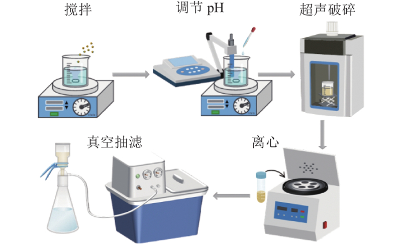
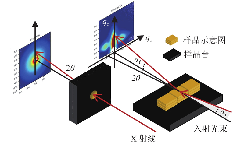
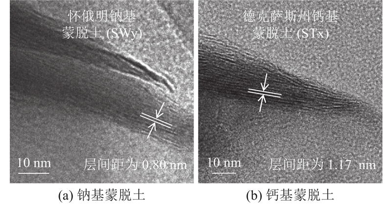
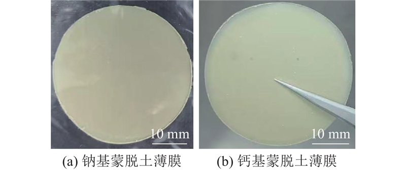
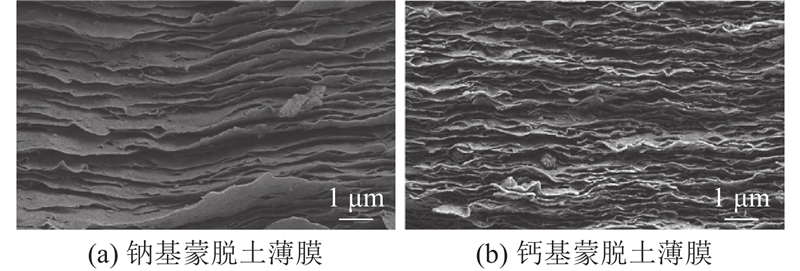
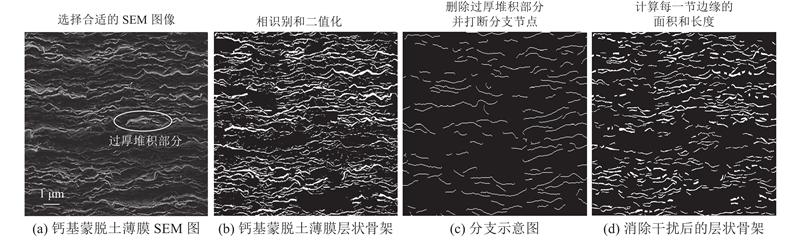
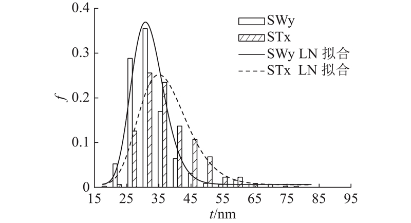
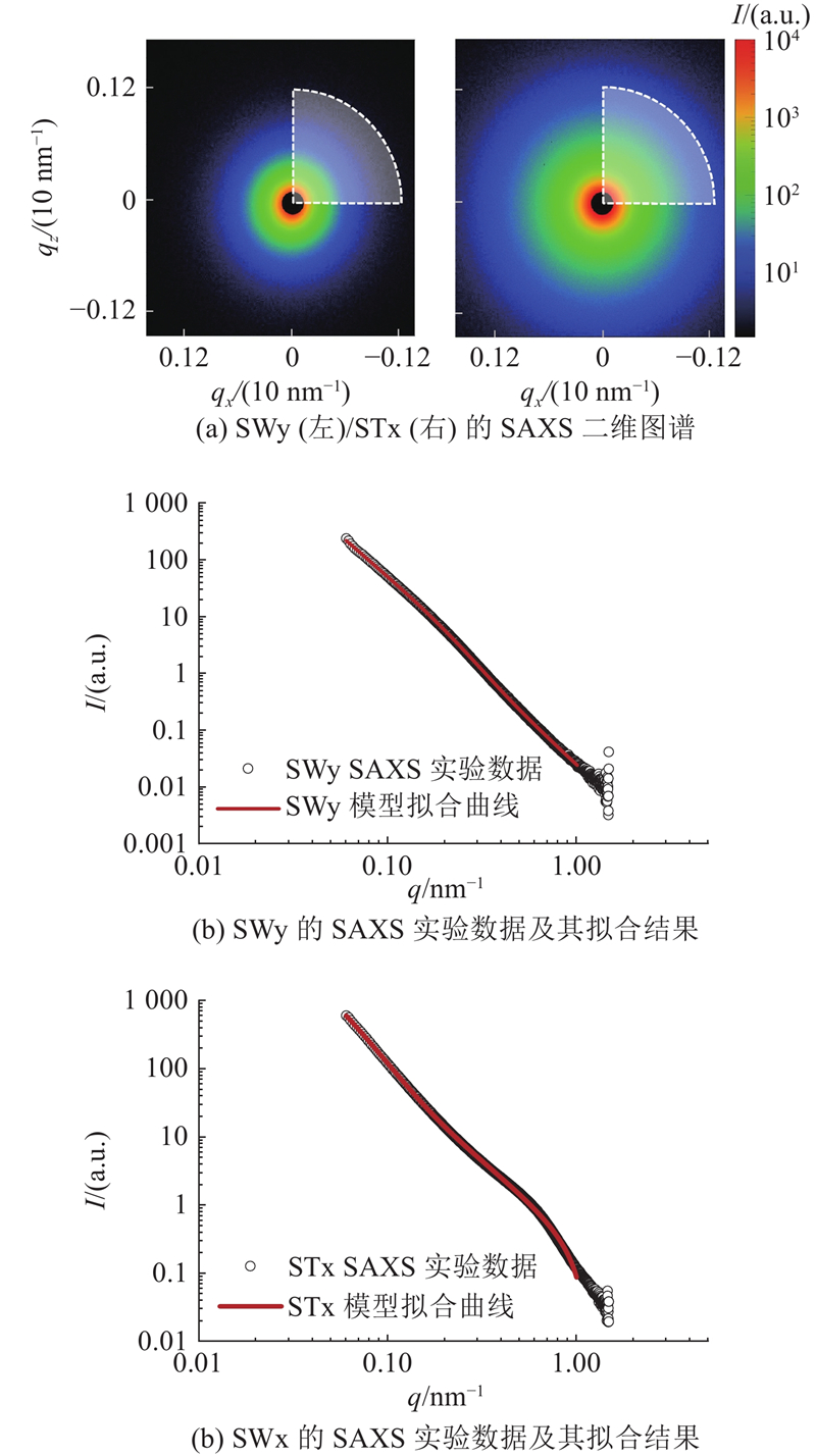
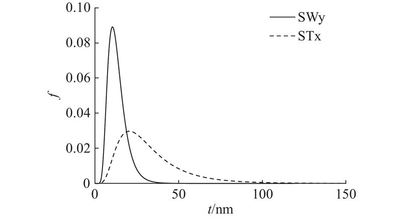
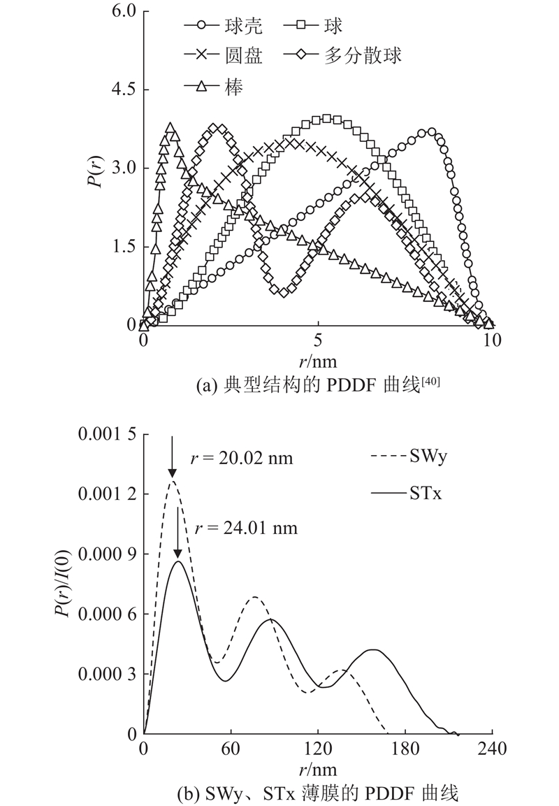
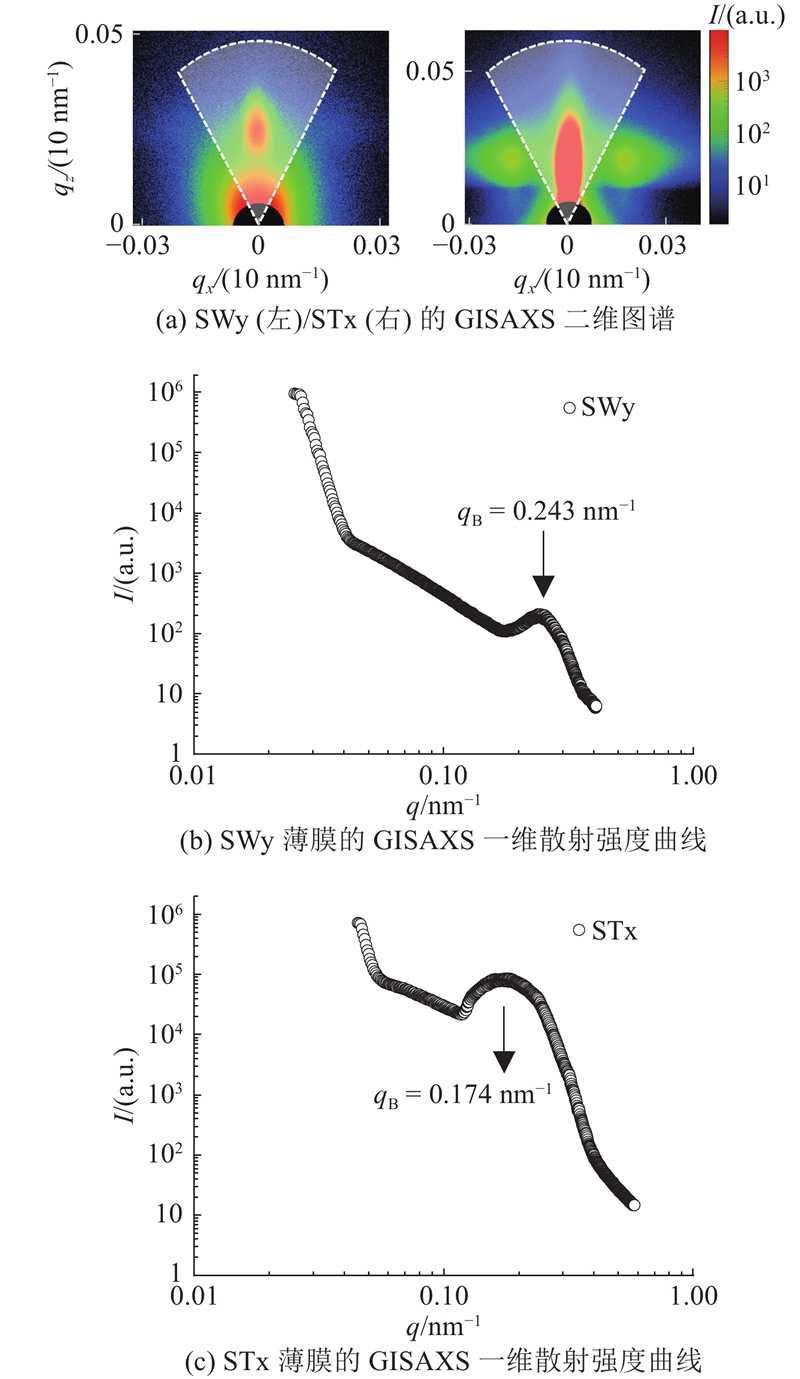
Abstract Quantitative analysis was performed on the lamellar thickness and spacing of montmorillonite aggregates in the sodium-based montmorillonite (SWy) and calcium-based montmorillonite (STx) film samples which were prepared via vacuum filtration by combining the scanning electron microscopy (SEM), small-angle X-ray scattering (SAXS) and grazing-incidence small-angle X-ray scattering (GISAXS) techniques and using the methods of SEM image recognition and scattering model fitting, to address the issue that existing characterization techniques struggle to balance the characterization of nanoscale local morphology and the analysis of macroscopic statistical properties. The results demonstrated that the montmorillonite aggregates in SWy films exhibited smaller average lamellar thickness (SEM: 33.73 nm; SAXS: 13.19 nm), with loosely stacked lamellar structures and significant interlayer porosity. The aggregates in STx films formed denser structures due to the ionic bridging effect of Ca2+, showing larger lamellar thickness (SEM: 39.23 nm; SAXS: 32.79 nm) and higher dispersion. The combination of SEM-based local morphology analysis, SAXS-based global statistics analysis, and GISAXS-based periodicity analysis enabled the quantitative characterization of feature sizes of the lamellar structures in montmorillonite films and the mutual verification of data reliability. The analysis results demonstrated the feasibility of this combined method in providing technical support for the directional design of functional montmorillonite films by integrating multi-scale structural information.
|
|
Received: 15 March 2024
Published: 15 December 2025
|
|
|
| Fund: 国家自然科学基金资助项目(42277178, 51779254). |
|
Corresponding Authors:
Yi DONG
E-mail: penglanyue22@mails.ucas.ac.cn;ydong@whrsm.ac.cn
|
蒙脱土薄膜层状微观结构表征
针对现有表征技术难以兼顾纳米级局部形貌表征与宏观统计特性分析的问题,采用真空抽滤法制备钠基蒙脱土(SWy)与钙基蒙脱土(STx)薄膜样品,通过联用扫描电镜(SEM)、小角X射线散射(SAXS)和掠入式小角X射线散射(GISAXS)技术,运用SEM图像识别与散射模型拟合方法,对蒙脱土类聚体的层厚和间距开展定量解析. 结果表明,SWy薄膜类聚体的平均层厚更小(SEM: 33.73 nm; SAXS: 13.19 nm),呈松散层状堆叠,层间孔隙显著. STx薄膜中的类聚体因Ca2+的离子桥联效应形成更致密的结构,平均层厚更大(SEM: 39.23 nm; SAXS: 32.79 nm),离散性更高. SEM局部形貌分析、SAXS全局统计分析及GISAXS周期性分析的联用实现了蒙脱土薄膜层状结构特征尺寸的定量表征,且能够互相验证数据的可靠性. 分析结果验证了该联用方法通过融合多尺度结构信息为蒙脱土功能薄膜定向设计提供技术支撑的可行性.
关键词:
蒙脱土薄膜,
层状结构,
微观结构表征,
小角X射线散射,
掠入式小角X射线散射
|
|
| [1] |
KOGURE T Visualization of clay minerals at the atomic scale[J]. Clay Minerals, 2020, 55 (3): 203- 218
doi: 10.1180/clm.2020.27
|
|
|
| [2] |
LAI Y H, CHIU C W, CHEN J G, et al Enhancing the performance of dye-sensitized solar cells by incorporating nanosilicate platelets in gel electrolyte[J]. Solar Energy Materials and Solar Cells, 2009, 93 (10): 1860- 1864
doi: 10.1016/j.solmat.2009.06.027
|
|
|
| [3] |
汪云逸, 邹楚文, 尹冉, 等 壳聚糖增强纳米纤维素-蒙脱土复合膜的结构与性能[J]. 复合材料学报, 2024, 41 (8): 4299- 4309
WANG Yunyi, ZOU Chuwen, YIN Ran, et al Structure and properties of chitosan enhanced cellulose nanofiber-montmorillonite composite membrane[J]. Acta Materiae Compositae Sinica, 2024, 41 (8): 4299- 4309
|
|
|
| [4] |
陈万锋, 胡鸿韬, 董良, 等 蒙脱土纳米片增强的仿生结构强韧水凝胶薄膜[J]. 化学通报, 2024, 87 (6): 710- 719
CHEN Wanfeng, HU Hongtao, DONG Liang, et al Montmorillonite nanosheet reinforced biomimetic structure strength tough hydrogel[J]. Chemistry, 2024, 87 (6): 710- 719
|
|
|
| [5] |
EBINA T, ISHII R, AIZAWA T, et al Development of clay-based film and its application to gas barrier layers of composite tanks[J]. Journal of the Japan Petroleum Institute, 2017, 60 (3): 121- 126
doi: 10.1627/jpi.60.121
|
|
|
| [6] |
YOSHIDA H, ARAI K, SUZUKI A, et al Development of a gas permeation measuring device and the evaluation of gas barrier property of clay-polymer nanocomposite films[J]. Clay Science, 2018, 22 (4): 95- 102
|
|
|
| [7] |
WANG Y C, HUANG T K, TUNG S H, et al Self-assembled clay films with a platelet–void multilayered nanostructure and flame-blocking properties[J]. Scientific Reports, 2013, 3: 2621
doi: 10.1038/srep02621
|
|
|
| [8] |
LIU M L, HUANG M, TIAN L Y, et al Two-dimensional nanochannel arrays based on flexible montmorillonite membranes[J]. ACS Applied Materials & Interfaces, 2018, 10 (51): 44915- 44923
|
|
|
| [9] |
SHAO J J, RAIDONGIA K, KOLTONOW A R, et al Self-assembled two-dimensional nanofluidic proton channels with high thermal stability[J]. Nature Communications, 2015, 6: 7602
doi: 10.1038/ncomms8602
|
|
|
| [10] |
ATANASOVA M T, FOCKE W W, LOOTS T Self-assembled rectorite films with remarkable mechanical performance: preparation, structural characterization, and properties[J]. Journal of Materials Science: Materials in Engineering, 2024, 19 (1): 17
doi: 10.1186/s40712-024-00161-z
|
|
|
| [11] |
MORGAN A B, GILMAN J W Characterization of polymer-layered silicate (clay) nanocomposites by transmission electron microscopy and X-ray diffraction: a comparative study[J]. Journal of Applied Polymer Science, 2003, 87 (8): 1329- 1338
doi: 10.1002/app.11884
|
|
|
| [12] |
STEFANESCU E A, DUNDIGALLA A, FERREIRO V, et al Supramolecular structures in nanocomposite multilayered films[J]. Physical Chemistry Chemical Physics, 2006, 8 (14): 1739- 1746
doi: 10.1039/b517880k
|
|
|
| [13] |
HAN J T, JANG J I, KIM H, et al Extremely efficient liquid exfoliation and dispersion of layered materials by unusual acoustic cavitation[J]. Scientific Reports, 2014, 4: 5133
doi: 10.1038/srep05133
|
|
|
| [14] |
PODSIADLO P, KAUSHIK A K, ARRUDA E M, et al Ultrastrong and stiff layered polymer nanocomposites[J]. Science, 2007, 318 (5847): 80- 83
doi: 10.1126/science.1143176
|
|
|
| [15] |
WONG M, ISHIGE R, WHITE K L, et al Large-scale self-assembled zirconium phosphate smectic layers via a simple spray-coating process[J]. Nature Communications, 2014, 5: 3589
doi: 10.1038/ncomms4589
|
|
|
| [16] |
MYLES A, GRIFFITH A, RIYAD M F, et al 3D-printed ceramics with aligned micro-platelets[J]. ACS Applied Engineering Materials, 2023, 1 (7): 1892- 1902
doi: 10.1021/acsaenm.3c00223
|
|
|
| [17] |
SAHA K, DEKA J, GOGOI R K, et al Applications of lamellar membranes reconstructed from clay mineral-based nanosheets: a review[J]. ACS Applied Nano Materials, 2022, 5 (11): 15972- 15999
doi: 10.1021/acsanm.1c03207
|
|
|
| [18] |
UMEMURA Y. Preparation and application of clay mineral films [M]// Developments in clay science. Amsterdam: Elsevier, 2018: 377–396.
|
|
|
| [19] |
DOR M, LEVI-KALISMAN Y, DAY-STIRRAT R J, et al Assembly of clay mineral platelets, tactoids, and aggregates: effect of mineral structure and solution salinity[J]. Journal of Colloid and Interface Science, 2020, 566: 163- 170
doi: 10.1016/j.jcis.2020.01.084
|
|
|
| [20] |
CHIU C W, LIN J J Self-assembly behavior of polymer-assisted clays[J]. Progress in Polymer Science, 2012, 37 (3): 406- 444
doi: 10.1016/j.progpolymsci.2011.07.007
|
|
|
| [21] |
WANG Y C, LIN J J Clay films with variable metal ions and self-assembled silicate layer-void nanostructures[J]. RSC Advances, 2014, 4 (12): 6356
doi: 10.1039/c3ra46628k
|
|
|
| [22] |
NAM H J, EBINA T, MIZUKAMI F Formability and properties of self-standing clay film by montmorillonite with different interlayer cations[J]. Colloids and Surfaces A: Physicochemical and Engineering Aspects, 2009, 346 (1/2/3): 158- 163
|
|
|
| [23] |
DAS P, MANNA S, BEHERA A K, et al Current synthesis and characterization techniques for clay-based polymer nano-composites and its biomedical applications: a review[J]. Environmental Research, 2022, 212: 113534
doi: 10.1016/j.envres.2022.113534
|
|
|
| [24] |
WALLEY P, ZHANG Y, EVANS J G Self-assembly of montmorillonite platelets during drying[J]. Bioinspiration & Biomimetics, 2012, 7 (4): 046004
|
|
|
| [25] |
郝文莉, 陈喜清, 屯妮萨·麦提赛伊迪, 等 立方体型水滑石/蒙脱土基复合材料的制备及其在PBAT中的应用[J]. 化工新型材料, 2022, 50 (4): 273- 277
HAO Wenli, CHEN Xiqing, TUNNISA Maitisaiyidi, et al Preparation of cubical hydrotalcite/montmorillonite composite and its application in PBAT[J]. New Chemical Materials, 2022, 50 (4): 273- 277
|
|
|
| [26] |
付英英, 李红轩, 吉利, 等 CrN和CrAlN薄膜的微观结构及在不同介质中的摩擦学性能[J]. 中国表面工程, 2012, 25 (6): 34- 41
FU Yingying, LI Hongxuan, JI Li, et al Microstructure and tribological properties of CrN and CrAlN films under different contact conditions[J]. China Surface Engineering, 2012, 25 (6): 34- 41
doi: 10.3969/j.issn.1007-9289.2012.06.006
|
|
|
| [27] |
杨子芹, 刘卫卫, 杨小兵, 等 蒙脱土有机改性对丁基橡胶复合材料微观结构与性能的影响[J]. 高分子材料科学与工程, 2011, 27 (9): 52- 55
YANG Ziqin, LIU Weiwei, YANG Xiaobing, et al Influence of montmorillonite organic modification on the microstructure and properties of IIR composites[J]. Polymer Materials Science & Engineering, 2011, 27 (9): 52- 55
|
|
|
| [28] |
安书香. 插层改性蒙脱土/木薯淀粉复合薄膜的制备及阻隔机理研究[D]. 南宁: 广西大学, 2019.
AN Shuxiang. Preparation and barrier mechanism of intercalated modified montmorillonite/tapioca starch composite film [D]. Nanning: Guangxi University, 2019.
|
|
|
| [29] |
LI P, WHITE K L, LIN C H, et al Mechanical reinforcement of epoxy with self-assembled synthetic clay in smectic order[J]. ACS Applied Materials & Interfaces, 2014, 6 (13): 10188- 10195
|
|
|
| [30] |
梁志扬 BOPP微孔薄膜孔径测试方法的研究[J]. 制造业自动化, 2020, 42 (7): 128- 130
LIANG Zhiyang The measurement method of the aperture of BOPP microporous film is briefly discussed[J]. Manufacturing Automation, 2020, 42 (7): 128- 130
|
|
|
| [31] |
VYDELINGUM S, LEVITZ P, MICHOT L J, et al Clay platelet orientation inside self-standing beidellite clay films: effect of silica nanospheres and link with macroscopic mechanical resistance[J]. Applied Clay Science, 2023, 231: 106740
doi: 10.1016/j.clay.2022.106740
|
|
|
| [32] |
SONG P, LI Q, ALMÁSY L, et al Fractionation of clay colloids and their synthetic utility in vanadium hydroxide-clay thin film formation[J]. Applied Surface Science, 2019, 481: 92- 98
doi: 10.1016/j.apsusc.2019.03.083
|
|
|
| [33] |
刘晓旭, 殷景华, 程伟东, 等 利用小角X射线散射技术研究组分对聚酰亚胺/Al2O3杂化薄膜界面特性与分形特征的影响[J]. 物理学报, 2011, 60 (5): 517- 522
LIU Xiaoxu, YIN Jinghua, CHENG Weidong, et al Research on interface and fractal characteristics of PI/Al2O3 films by SAXS[J]. Acta Physica Sinica, 2011, 60 (5): 517- 522
doi: 10.7498/aps.60.056101
|
|
|
| [34] |
SEMERARO E F, HENGL N, KARROUCH M, et al Layered organization of anisometric cellulose nanocrystals and beidellite clay particles accumulated near the membrane surface during cross-flow ultrafiltration: in situ SAXS and ex situ SEM/WAXD characterization[J]. Colloids and Surfaces A: Physicochemical and Engineering Aspects, 2020, 584: 124030
doi: 10.1016/j.colsurfa.2019.124030
|
|
|
| [35] |
BAKER J L, JIMISON L H, MANNSFELD S, et al Quantification of thin film crystallographic orientation using X-ray diffraction with an area detector[J]. Langmuir, 2010, 26 (11): 9146- 9151
doi: 10.1021/la904840q
|
|
|
| [36] |
LENG Y, LI Q, TIAN Q, et al (Ce-Al)-oxide pillared bentonite: a high affinity sorbent for plutonium[J]. Journal of Hazardous Materials, 2018, 352: 121- 129
doi: 10.1016/j.jhazmat.2018.03.028
|
|
|
| [37] |
CHANG J, SHAO H, LIU B, et al Control of nanostructures through pH-dependent self-assembly of nanoplatelets[J]. Journal of Colloid and Interface Science, 2021, 582: 439- 445
doi: 10.1016/j.jcis.2020.07.093
|
|
|
| [38] |
张云龙, 项伟, 黄伟, 等 钠基蒙脱土水合演化机制[J]. 岩土力学, 2019, 40 (11): 4391- 4400
ZHANG Yunlong, XIANG Wei, HUANG Wei, et al Hydration evolution mechanism of sodium montmorillonite[J]. Rock and Soil Mechanics, 2019, 40 (11): 4391- 4400
|
|
|
| [39] |
李昆鹏, 陈永贵, 叶为民, 等 高压实膨润土孔隙结构特征研究进展[J]. 岩土工程学报, 2022, 44 (3): 399- 408
LI Kunpeng, CHEN Yonggui, YE Weimin, et al Advances in studies on pore structure of highly compacted bentonite[J]. Chinese Journal of Geotechnical Engineering, 2022, 44 (3): 399- 408
doi: 10.11779/CJGE202203001
|
|
|
| [40] |
陈孔磊. 高庙子膨润土中金属阳离子的空间分布规律及吸附特性研究[D]. 桂林: 桂林理工大学, 2022.
CHEN Konglei. A study on the spatial distribution and adsorptive behavior of metallic ions in Gaomiaozi bentonite [D]. Guilin: Guilin University of Technology, 2022.
|
|
|
| [41] |
GLATTER O. Modern methods of data analysis in small-angle scattering and light scattering [M]// BRUMBERGER H. Modern aspects of small-angle scattering. Dordrecht: Springer, 1995: 107–180.
|
|
|
| [42] |
WU D Q, CHU B, LUNDBERG R D, et al Small-angle X-ray scattering (SAXS) studies of sulfonated polystyrene ionomers. 2. Correlation function analysis[J]. Macromolecules, 1993, 26 (5): 1000- 1007
doi: 10.1021/ma00057a019
|
|
|
| [43] |
SVERGUN D I, KOCH M H J Small-angle scattering studies of biological macromolecules in solution[J]. Reports on Progress in Physics, 2003, 66 (10): 1735- 1782
doi: 10.1088/0034-4885/66/10/R05
|
|
|
|
Viewed |
|
|
|
Full text
|
|
|
|
|
Abstract
|
|
|
|
|
Cited |
|
|
|
|
| |
Shared |
|
|
|
|
| |
Discussed |
|
|
|
|