| Animal sciences & veterinary medicines |
|

|
|
|
| Preparation of polyclonal antibodies and subcellular localization of non-structural protein 3 encoded by feline coronavirus |
Ziyi WANG1( ),Zi’an JIN1,2,Chenhe LU1,Zhi QIAO3,Shengwen WANG1,Yan YAN1,Jiyong ZHOU1,Xiaojuan ZHENG1() ),Zi’an JIN1,2,Chenhe LU1,Zhi QIAO3,Shengwen WANG1,Yan YAN1,Jiyong ZHOU1,Xiaojuan ZHENG1() |
1.Key Laboratory of Animal Virology, Ministry of Agriculture and Rural Affairs, Center for Veterinary Sciences, Zhejiang University, Hangzhou 310058, Zhejiang, China
2.Hainan Institute of Zhejiang University, Sanya 572025, Hainan, China
3.ZJU-Partner Joint Research Center, Hangzhou 310058, Zhejiang, China |
|
|
|
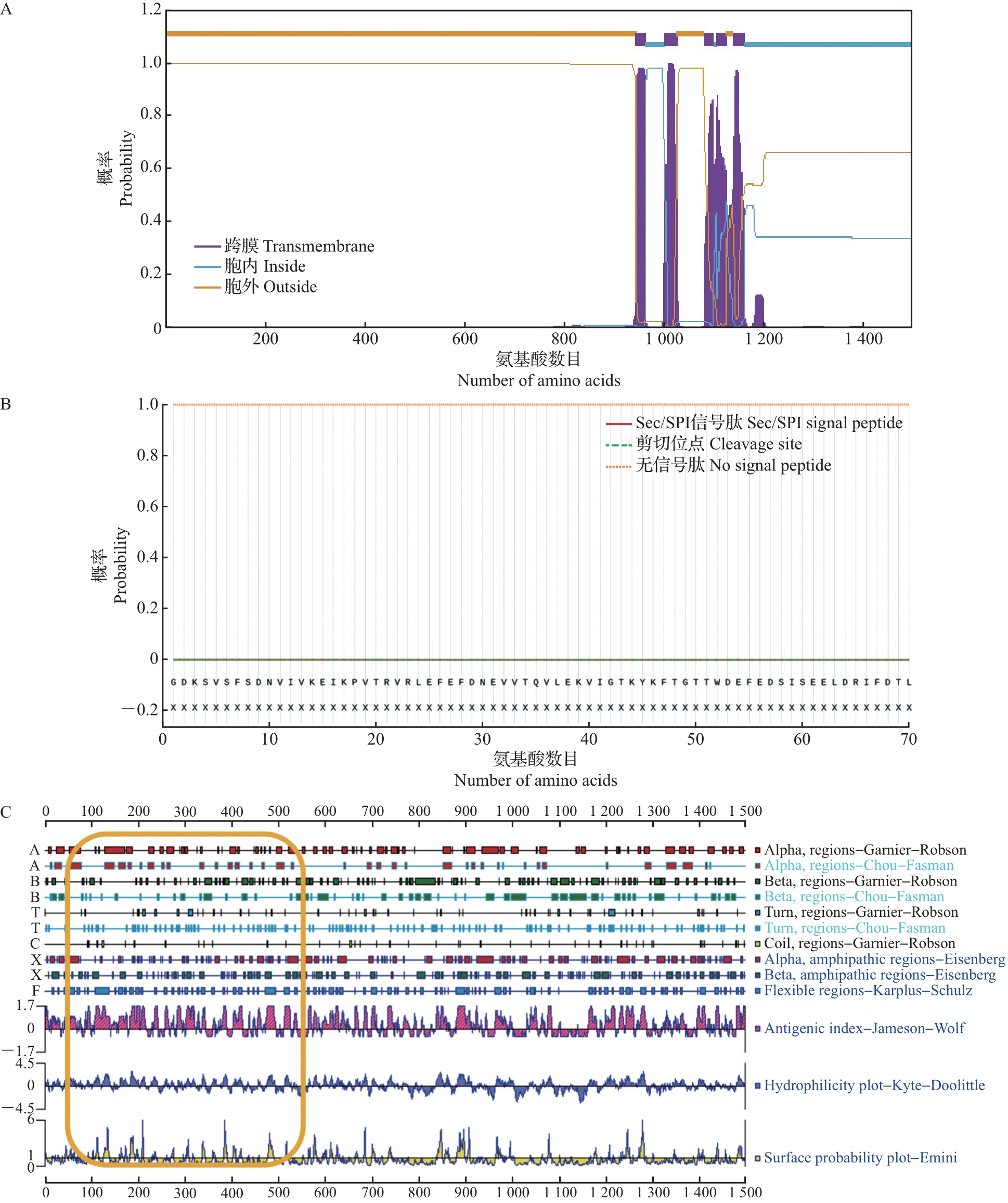
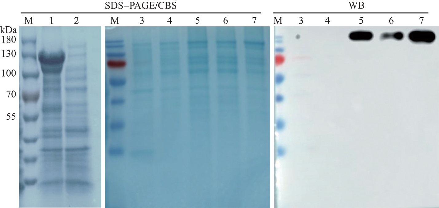
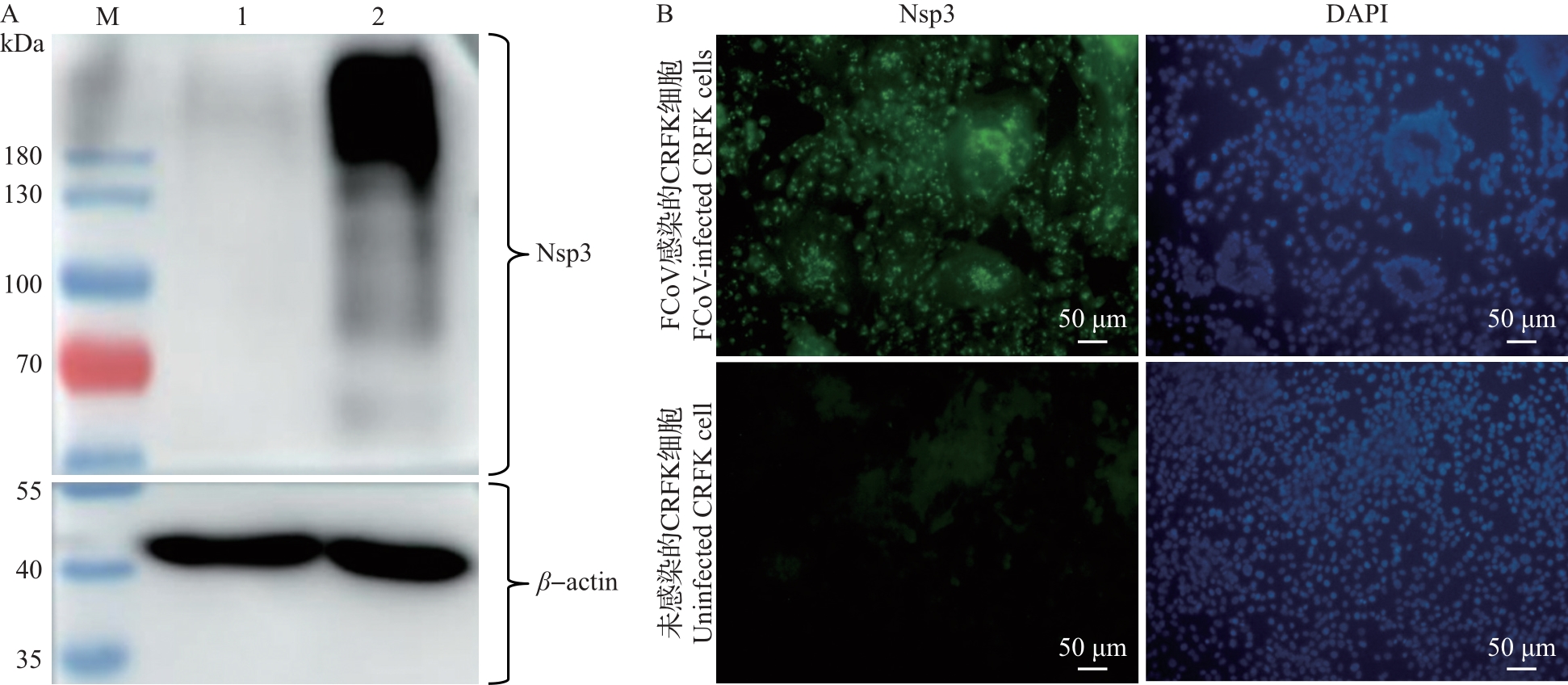
Abstract The non-structural protein 3 (Nsp3) of coronavirus, a component of the replication and transcription complex, is one of the potentially important antiviral targets. In this study, the transmembrane region, signal peptide, and epitope of Nsp3 were predicted, and then the region with better antigenicity (50-550 amino acids) of Nsp3 protein in a representative strain (WSU 79-1683) of type Ⅱ feline coronavirus (FCoV) was amplified by polymerase chain reaction. Subsequently, it was subcloned into pCOLD-TF prokaryotic expression vector. Under the low-temperature condition, the recombinant fusion protein His-Nsp3 with a molecular weight of about 130 kDa was successfully induced by isopropylthio-β-D-galactoside. The targeted recombinant protein His-Nsp3 was purified using a non-denaturing nickel affinity column, and the purified protein was used as an antigen to immunize BALB/c mice for preparing Nsp3 polyclonal antiserum. Western blotting (WB) and indirect immunofluorescence assay (IFA) results showed that Nsp3 polyclonal antiserum could specifically recognize Nsp3 protein in FCoV-infected cells. The subcellular localization of Nsp3 protein in FCoV-infected cells was studied by double-labeling IFA combined with laser confocal microscopy. The results showed that Nsp3 protein aggregated in FCoV-infected cells and co-localized with the endoplasmic reticulum. The specific antibody preparation and subcellular localization study of Nsp3 protein provided an important basis for further analysis of the biological function of Nsp3 protein.
|
|
Received: 01 August 2022
Published: 25 October 2023
|
|
|
|
Corresponding Authors:
Xiaojuan ZHENG
E-mail: 21917102@zju.edu.cn;zhengxiaojuan@zju.edu.cn
|
猫冠状病毒非结构蛋白3的多克隆抗体制备及亚细胞定位
冠状病毒的非结构蛋白3(non-structural protein 3, Nsp3)是复制/转录复合体的组成部分,也是重要的潜在抗病毒靶标之一。本研究在对Nsp3进行跨膜区、信号肽和抗原表位预测的基础上,通过聚合酶链反应扩增Ⅱ型猫冠状病毒(feline coronavirus, FCoV)代表毒株(WSU 79-1683)Nsp3蛋白中抗原性较好的区段(第50—550位氨基酸),随后将其亚克隆到pCOLD-TF原核表达载体上。在低温条件下,经异丙基硫代-β-D-半乳糖苷诱导,成功表达分子量约为130 kDa的His-Nsp3重组融合蛋白。在非变性条件下,采用镍柱亲和层析获得了纯度较高的目的重组蛋白。以纯化的重组蛋白为抗原免疫BALB/c小鼠,制备Nsp3多克隆抗血清(多抗血清),蛋白质印迹法(Western blotting, WB)和间接免疫荧光试验(indirect immunofluorescence assay, IFA)结果显示,Nsp3多抗血清能特异性识别FCoV感染细胞中的Nsp3蛋白。采用免疫荧光双标法结合激光共聚焦显微镜对FCoV感染细胞中Nsp3蛋白的亚细胞定位进行分析,结果发现,FCoV感染细胞中Nsp3发生聚集现象,并与内质网存在共定位现象。Nsp3蛋白的特异性抗体制备以及亚细胞定位研究为进一步开展Nsp3蛋白的生物学功能分析奠定了基础。
关键词:
猫冠状病毒,
非结构蛋白3,
原核表达,
多克隆抗体,
亚细胞定位
|
|
| [1] |
ACAR D D, STROOBANTS V J E, FAVOREEL H, et al. Identification of peptide domains involved in the subcellular localization of the feline coronavirus 3b protein[J]. Journal of General Virology, 2019, 100(10): 1417-1430. DOI: 10.1099/jgv.0.001321
doi: 10.1099/jgv.0.001321
|
|
|
| [2] |
GONZÁLEZ J M, GOMEZ-PUERTAS P, CAVANAGH D, et al. A comparative sequence analysis to revise the current taxonomy of the family Coronaviridae [J]. Archives of Virology, 2003, 148(11): 2207-2235. DOI: 10.1007/s00705-003-0162-1
doi: 10.1007/s00705-003-0162-1
|
|
|
| [3] |
KIPAR A, MELI M L. Feline infectious peritonitis: Still an enigma?[J]. Veterinary Pathology, 2014, 51(2): 505-526. DOI: 10.1177/0300985814522077
doi: 10.1177/0300985814522077
|
|
|
| [4] |
KENNEDY M A. Feline infectious peritonitis: update on pathogenesis, diagnostics, and treatment[J]. Veterinary Clinics of North America: Small Animal Practice, 2020, 50(5): 1001-1011. DOI: 10.1016/j.cvsm.2020.05.002
doi: 10.1016/j.cvsm.2020.05.002
|
|
|
| [5] |
PEDERSEN N C. A review of feline infectious peritonitis virus infection: 1963—2008[J]. Journal of Feline Medicine and Surgery, 2009, 11(4): 225-258. DOI: 10.1016/j.jfms.2008.09.008
doi: 10.1016/j.jfms.2008.09.008
|
|
|
| [6] |
ROTTIER P J M. The molecular dynamics of feline corona-viruses[J]. Veterinary Microbiology, 1999, 69(1/2): 117-125.
|
|
|
| [7] |
DRECHSLER Y, VASCONCELOS E J R, GRIGGS L M, et al. Host gene expression of macrophages in response to feline coronavirus infection[J]. Cells, 2020, 9(6): 1431. DOI: 10.3390/cells9061431
doi: 10.3390/cells9061431
|
|
|
| [8] |
NEUMAN B W. Bioinformatics and functional analyses of coronavirus nonstructural proteins involved in the formation of replicative organelles[J]. Antiviral Research, 2016, 135: 97-107. DOI: 10.1016/j.antiviral.2016.10.005
doi: 10.1016/j.antiviral.2016.10.005
|
|
|
| [9] |
ROHAIM M A, NAGGAR R, CLAYTON E, et al. Struc-tural and functional insights into non-structural proteins of coronaviruses[J]. Microbial Pathogenesis, 2020, 150(12): 104641. DOI: 10.1016/j.micpath.2020.104641
doi: 10.1016/j.micpath.2020.104641
|
|
|
| [10] |
JIANG Y L, TONG K J, YAO R B, et al. Genome-wide analysis of protein-protein interactions and involvement of viral proteins in SARS-CoV-2 replication[J]. Cell & Bio-science, 2021, 11: 140. DOI: 10.1186/s13578-021-00644-y
doi: 10.1186/s13578-021-00644-y
|
|
|
| [11] |
OOSTRA M, HAGEMEIJER M C, VAN GENT M, et al. Topology and membrane anchoring of the coronavirus replication complex: not all hydrophobic domains of nsp3 and nsp6 are membrane spanning[J]. Journal of Virology, 2008, 82(24): 12392-12405. DOI: 10.1128/JVI.01219-08
doi: 10.1128/JVI.01219-08
|
|
|
| [12] |
HURST K R, KOETZNER C A, MASTERS P S. Characterization of a critical interaction between the corona-virus nucleocapsid protein and nonstructural protein 3 of the viral peplicase-transcriptase complex[J]. Journal of Virology, 2013, 87(16): 9159-9172. DOI: 10.1128/JVI.01275-13
doi: 10.1128/JVI.01275-13
|
|
|
| [13] |
MATTHES N, MESTERS J R, COUTARD B, et al. The non-structural protein Nsp10 of mouse hepatitis virus binds zinc ions and nucleic acids[J]. FEBS Letters, 2006, 580(17): 4143-4149. DOI: 10.1016/j.febslet.2006.06.061
doi: 10.1016/j.febslet.2006.06.061
|
|
|
| [14] |
DONALDSON E F, GRAHAM R L, SIMS A C, et al. Analysis of murine hepatitis virus strain A59 temperature-sensitive mutant TS-LA6 suggests that nsp10 plays a critical role in polyprotein processing[J]. Journal of Virology, 2007, 81(13): 7086-7098. DOI: 10.1128/JVI.00049-07
doi: 10.1128/JVI.00049-07
|
|
|
| [15] |
DRECHSLER Y, DVM A A, DVM F J B, et al. Feline coronavirus in multicat environments[J]. Veterinary Clinics of North America: Small Animal Practice, 2011, 41(6): 1133-1169. DOI: 10.1016/j.cvsm.2011.08.004
doi: 10.1016/j.cvsm.2011.08.004
|
|
|
| [16] |
STOKES H L, BALIJI S, HUI C G, et al. A new cistron in the murine hepatitis virus replicase gene[J]. Journal of Virology, 2010, 84(19): 10148-10158. DOI: 10.1128/JVI.00901-10
doi: 10.1128/JVI.00901-10
|
|
|
| [17] |
RAJPOOT S, ALAGUMUTHU M, BAIG M S. Dual targeting of 3CLpro and PLpro of SARS-CoV-2: a novel structure-based design approach to treat COVID-19[J]. Current Research in Structural Biology, 2021, 3: 9-18. DOI: 10.1016/j.crstbi.2020.12.001
doi: 10.1016/j.crstbi.2020.12.001
|
|
|
| [18] |
JAIMES J A, MILLET J K, STOUT A E, et al. A tale of two viruses: the distinct spike glycoproteins of feline coronaviruses[J]. Viruses, 2020, 12(1): 83. DOI: 10.3390/v12010083
doi: 10.3390/v12010083
|
|
|
| [19] |
LIN C, CHAN K R, OOI E E, et al. Animal coronavirus diseases: parallels with COVID-19 in humans[J]. Viruses, 2021, 13(8): 1507. DOI: 10.3390/v13081507
doi: 10.3390/v13081507
|
|
|
| [20] |
PALTRINIERI S, ROSSI G, GIORDANO A. Relationship between rate of infection and markers of inflammation/immunity in Holy Birman cats with feline coronavirus[J]. Research in Veterinary Science, 2014, 97(2): 263-270. DOI: 10.1016/j.rvsc.2014.08.009
doi: 10.1016/j.rvsc.2014.08.009
|
|
|
| [21] |
蒋平平,王汀.新冠病毒的免疫性和新冠疫苗研发进展[J].中国药剂学杂志(网络版),2022,20(1):40-46. DOI:10.14146/j.cnki.cjp.2022.01.004
JIANG P P, WANG T. SARS-CoV-2 immunity and vaccine progress[J]. Chinese Journal of Pharmaceutics (Online Edition), 2022, 20(1): 40-46. (in Chinese with English abstract)
doi: 10.14146/j.cnki.cjp.2022.01.004
|
|
|
| [22] |
ISLAM A, FERDOUS J, ISLAM S, et al. Evolutionary dynamics and epidemiology of endemic and emerging coronaviruses in humans, domestic animals, and wildlife[J]. Viruses, 2021, 13(10): 1908. DOI: 10.3390/v13101908
doi: 10.3390/v13101908
|
|
|
| [23] |
SILVA C S, MULLIS L B, Jr, PEREIRA O, et al. Human respiratory coronaviruses detected in patients with influenza-like illness in Arkansas, USA[J]. Virology & Mycology, 2014, S2: 004. DOI: 10.4172/2161-0517.S2-004
doi: 10.4172/2161-0517.S2-004
|
|
|
| [24] |
HOSSAIN M G, JAVED A, AKTER S, et al. SARS-CoV-2 host diversity: an update of natural infections and experi-mental evidence[J]. Journal of Microbiology, Immunology and Infection, 2020, 54(2): 175-181. DOI: 10.1016/j.jmii.2020.06.006
doi: 10.1016/j.jmii.2020.06.006
|
|
|
| [25] |
FOLEY J E, POLAND A, CARLSON J, et al. Patterns of feline coronavirus infection and fecal shedding from cats in multiple-cat environments[J]. Journal of the American Veter-inary Medical Association, 1997, 210(9): 1307-1312.
|
|
|
| [26] |
PEDERSEN N C. An update on feline infectious peritonitis: virology and immunopathogenesis[J]. The Veterinary Journal, 2014, 201(2): 123-132. DOI: 10.1016/j.tvjl.2014.04.017
doi: 10.1016/j.tvjl.2014.04.017
|
|
|
| [27] |
JAIMES J A, WHITTAKER G R. Feline coronavirus: insights into viral pathogenesis based on the spike protein structure and function[J]. Virology, 2018, 517: 108-121. DOI: 10.1016/j.virol.2017.12.027
doi: 10.1016/j.virol.2017.12.027
|
|
|
| [28] |
YE G, WANG X W, TONG X H, et al. Structural basis for inhibiting porcine epidemic diarrhea virus replication with the 3C-like protease inhibitor GC376[J]. Viruses, 2020, 12(2): 240. DOI: 10.3390/v12020240
doi: 10.3390/v12020240
|
|
|
| [29] |
方馨悦,吴英理.新型冠状病毒木瓜样蛋白酶及其抑制剂研究进展[J].生命的化学,2021,41(8):1784-1795. DOI:10.13488/j.smhx.20210156
FANG X Y, WU Y L. Papain-like proteases of SARS-CoV-2 and its current inhibitors[J]. Chemistry of Life, 2021, 41(8): 1784-1795. (in Chinese with English abstract)
doi: 10.13488/j.smhx.20210156
|
|
|
|
Viewed |
|
|
|
Full text
|
|
|
|
|
Abstract
|
|
|
|
|
Cited |
|
|
|
|
| |
Shared |
|
|
|
|
| |
Discussed |
|
|
|
|