| Computer Technology, Information Engineering |
|


|
|
|
| Human hemorheology information evaluation based on Hilbert-Huang transform to decompose photoplethysmography signal |
Lu YU( ),Long-zhe JIN*(),Ming-wei XU,Jian-guo LIU ),Long-zhe JIN*(),Ming-wei XU,Jian-guo LIU |
| School of Civil and Resource Engineering, University of Science and Technology Beijing, Beijing 100083, China |
|
|
|
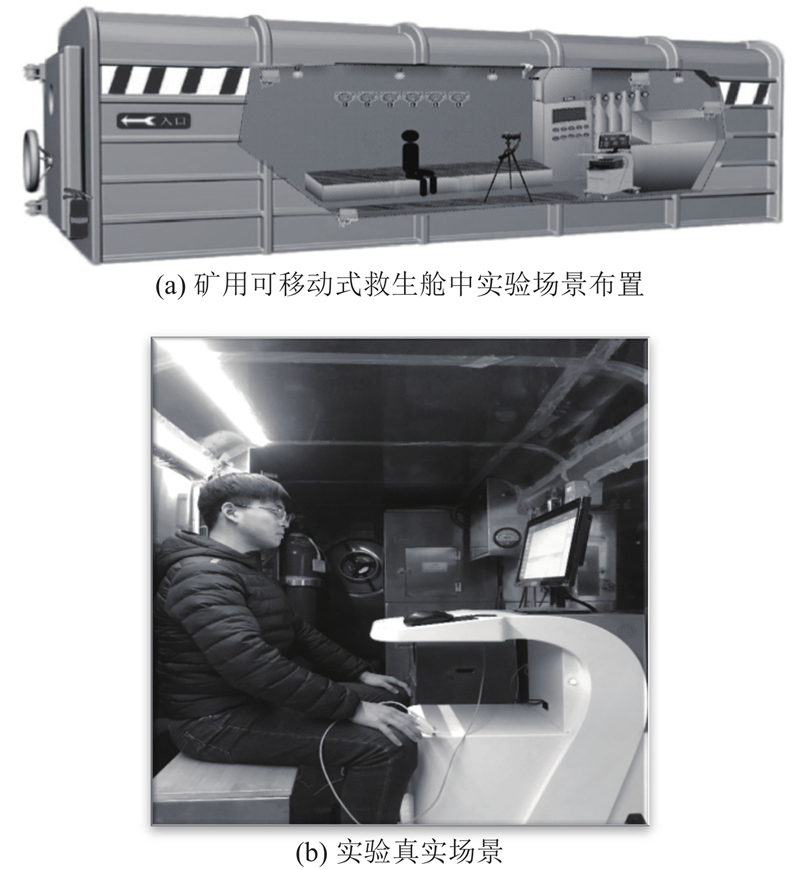
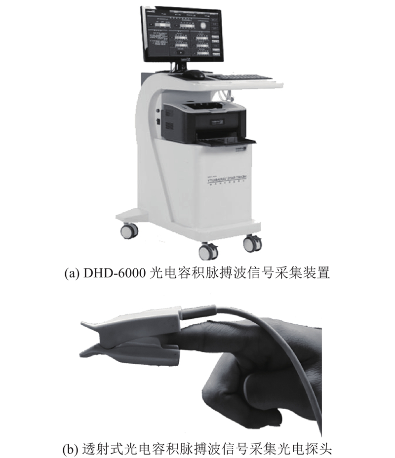
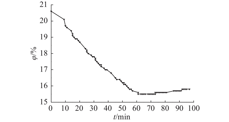
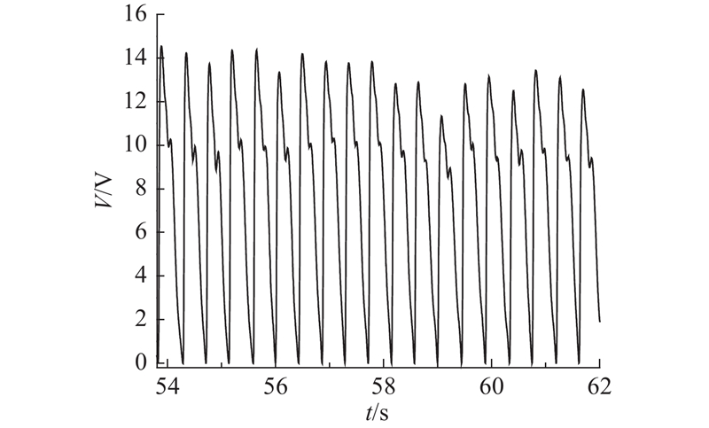
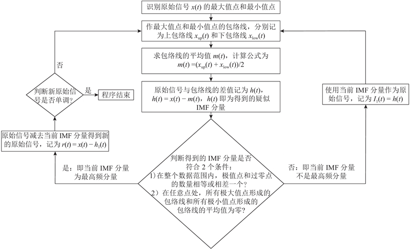
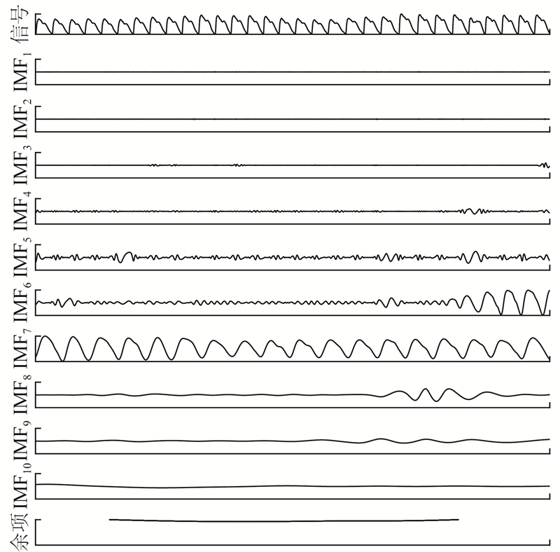
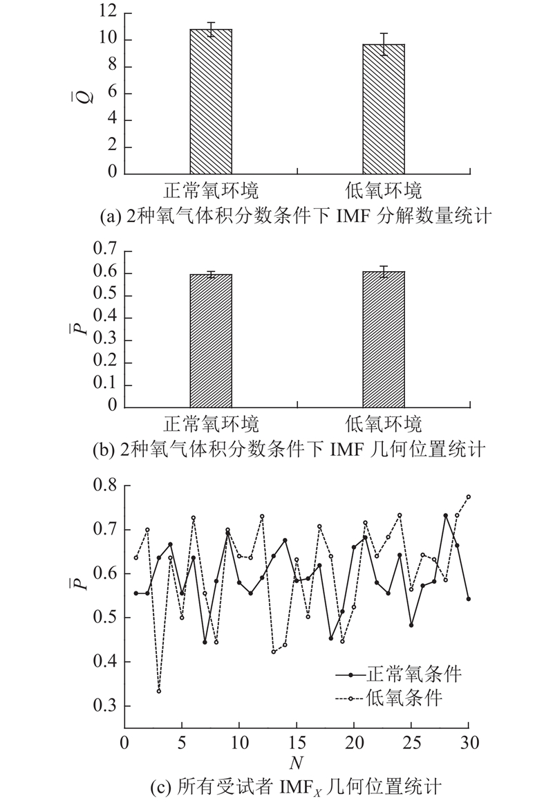
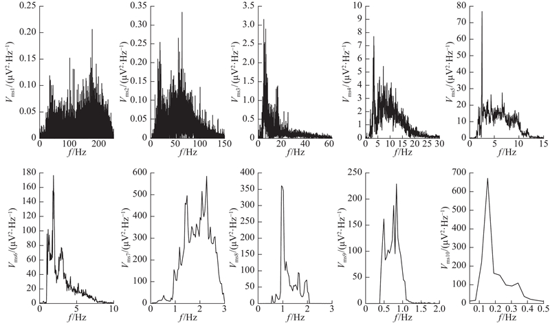
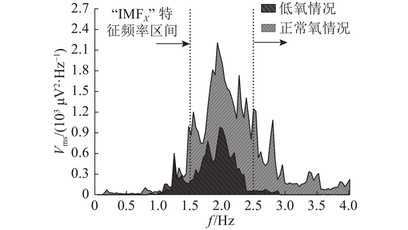
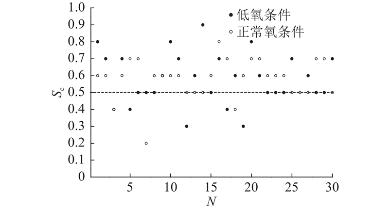
Abstract A hypoxia experiment was designed, in order to identify the dynamic component of the hemorheology information in the photoplethysmography (PPG) signal and analyze its characteristics. A total of thirty subjects were measured for PPG signals under normal oxygen volume fraction condition (20%~21%) and low oxygen volume fraction condition (15%~16%), respectively. The signal was analyzed based on the Hilbert-Huang transform (HHT) algorithm. The empirical mode decomposition results show that the dynamic component actually representing the hemorheology information of the PPG signal is intrinsic mode function IMFX. There are two time domain features of IMFX, one is a waveform similar to the arterial systolic relaxation, and the other is a periodic oscillation. The instantaneous frequency and marginal spectrum of IMFX were obtained based on the Hilbert transform algorithm, and the instantaneous frequency was mostly 1.5~2.5 Hz. In the hypoxic environment, the amplitude of the Hilbert marginal spectrum in the above frequency range is significantly smaller than that of the normal oxygen environment (P<0.05), which proves that this feature can be used to determine the hemorheological changes caused by hypoxia.
|
|
Received: 22 May 2019
Published: 10 March 2020
|
|
|
|
Corresponding Authors:
Long-zhe JIN
E-mail: yulubaobeihao@163.com;lzjin@ustb.edu.cn
|
基于HHT分解光电容积脉搏波信号的人体血液流变信息评估
为了识别人体光电容积脉搏波(PPG)信号中表征血液流变信息的动力分量并分析其特点,设计低氧实验. 测量30位受试者在正常氧(20%~21%)和极端低氧(15%~16%)2种氧气体积分数环境中的PPG信号,利用希尔伯特黄变换(HHT)算法分解信号. 通过经验模式分解得到,PPG信号中实际表征血液流变信息的动力分量为固有模式函数IMFX,其时域特点有2个,一个是有类似动脉收缩舒张的波形,另一个是周期性振荡. 基于Hilbert变换得到IMFX的瞬时频率和边际谱,其瞬时频率大多为1.5~2.5 Hz,且在低氧环境中此频率段内的边际谱幅值显著小于正常氧环境情况下的(P<0.05),证明利用该特征可以有效识别低氧诱导的血液流变变化.
关键词:
光电容积描记术(PPG),
希尔伯特黄变换(HHT),
固有模态函数,
经验模式分解,
血液流变学,
低氧环境
|
|
| [1] |
ALLEN J Photoplethysmography and its application in clinical physiological measurement[J]. Physiological Measurement, 2007, 28 (3): 1- 39
doi: 10.1088/0967-3334/28/3/R01
|
|
|
| [2] |
REISNER A, SHALTIS P A, MCCOMBIE D, et al Utility of the photoplethysmogram in circulatory monitoring[J]. Anesthesiology, 2008, 108 (5): 950- 958
doi: 10.1097/ALN.0b013e31816c89e1
|
|
|
| [3] |
KAMAL A A R, HARNESS J B, IRVING G, et al Skin photoplethysmography: a review[J]. Computer Methods and Programs in Biomedicine, 1989, 28 (4): 257- 269
doi: 10.1016/0169-2607(89)90159-4
|
|
|
| [4] |
KAMSHILIN A A, NIPPOLAINEN E, SIDOROV I S, et al A new look at the essence of the imaging photoplethysmography[J]. Scientific Reports, 2014, 5 (5): 10494
|
|
|
| [5] |
VOLKOV M V, MARGARYANTS N B, POTEMKIN A V, et al Video capillaroscopy clarifies mechanism of the photoplethysmographic waveform appearance[J]. Scientific Reports, 2017, 7 (1): 13298
doi: 10.1038/s41598-017-13552-4
|
|
|
| [6] |
NITZAN M, ADAR Y, HOFFMAN E, et al Comparison of systolic blood pressure values obtained by photoplethysmography and by korotkoff sounds[J]. Sensors, 2013, 13 (11): 14797- 14812
doi: 10.3390/s131114797
|
|
|
| [7] |
TAMURA T, MAEDA Y, SEKINE M, et al Wearable photoplethysmographic sensors: past and present[J]. Electronics, 2014, 3 (2): 282- 302
doi: 10.3390/electronics3020282
|
|
|
| [8] |
DALY S M, LEAHY M J ‘Go with the flow’: a review of methods and advancements in blood flow imaging[J]. Journal of Biophotonics, 2013, 6 (3): 217- 255
doi: 10.1002/jbio.201200071
|
|
|
| [9] |
WOWERN E V, ?STLING G, NILSSON P M, et al Digital photoplethysmography for assessment of arterial stiffness: repeatability and comparison with applanation tonometry[J]. Plos One, 2015, 10 (8): e0135659
doi: 10.1371/journal.pone.0135659
|
|
|
| [10] |
NJOUM H, KYRIACOU P A Photoplethysmography for the assessment of haemorheology[J]. Scientific Reports, 2017, 7 (1): 1406
doi: 10.1038/s41598-017-01636-0
|
|
|
| [11] |
LEE C, SIK S H, LEE M Relations between ac-dc components and optical path length in photoplethysmography[J]. Journal of Biomedical Optics, 2011, 16 (7): 077012
doi: 10.1117/1.3600769
|
|
|
| [12] |
KIM J M, CHOI J K, CHOI M, et al Assessment of cerebral autoregulation using continuous-wave near-infrared spectroscopy during squat-stand maneuvers in subjects with symptoms of orthostatic intolerance[J]. Scientific Reports, 2018, 8 (1): 13257
doi: 10.1038/s41598-018-31685-y
|
|
|
| [13] |
XING X, SUN M Optical blood pressure estimation with photoplethysmography and FFT-based neural networks[J]. Biomedical Optics Express, 2016, 8 (7): 3007- 3020
|
|
|
| [14] |
RAM M R, MADHAV K V, KRISHNA E H, et al A novel approach for motion artifact reduction in PPG signals based on AS-LMS adaptive filter[J]. IEEE Transactions on Instrumentation and Measurement, 2012, 61 (5): 1445- 1457
doi: 10.1109/TIM.2011.2175832
|
|
|
| [15] |
WANG C, ZHANG J L, XUN B W Monitoring heart and respiratory rates at radial artery based on PPG[J]. Optik, 2013, 19 (124): 3954- 3956
|
|
|
| [16] |
ELGENDI M Optimal signal quality index for photoplethysmogram signals[J]. Bioengineering, 2016, 4 (3): 21
|
|
|
| [17] |
于露, 金龙哲, 徐明伟, 等 基于光电容积脉搏波的有限空间生理疲劳测量[J]. 工程科学学报, 2018, 40 (10): 1215- 1222
YU Lu, JIN Long-zhe, XU Ming-wei, et al Confined space physiological fatigue measurement based on photoplethysmographypulse wave signal[J]. Chinese Journal of Engineering, 2018, 40 (10): 1215- 1222
|
|
|
| [18] |
HUANG N E, WU Z A review on Hilbert‐Huang transform: method and its applications to geophysical studies[J]. Reviews of Geophysics, 2008, 46 (2): 1- 23
|
|
|
| [19] |
PENG Z K, PETER W T, CHU F L A comparison study of improved Hilbert-Huang transform and wavelet transform: application to fault diagnosis for rolling bearing[J]. Mechanical Systems and Signal Processing, 2005, 19 (5): 974- 988
doi: 10.1016/j.ymssp.2004.01.006
|
|
|
| [20] |
YAN R Q, GAO R X Hilbert-Huang transform-based vibration signal analysis for machine health monitoring[J]. IEEE Transactions on Instrumentation and Measurement, 2006, 55 (6): 2320- 2329
doi: 10.1109/TIM.2006.887042
|
|
|
| [21] |
WU Z H, HUANG N E Ensemble empirical mode decomposition: a noise-assisted data analysis method[J]. Advances in Adaptive Data Analysis, 2009, 1 (01): 1- 41
doi: 10.1142/S1793536909000047
|
|
|
| [22] |
AYACHE S S, AL-ANI T, LEFAUCHEUR J P Distinction between essential and physiological tremor using Hilbert-Huang transform[J]. Neurophysiologie Clinique/Clinical Neurophysiology, 2014, 44 (2): 203- 212
|
|
|
| [23] |
TYAN C C, LIU S H, CHEN J Y, et al A novel noninvasive measurement technique for analyzing the pressure pulse waveform of the radial artery[J]. IEEE Transactions on Bio-medical Engineering, 2008, 55 (1): 288- 297
doi: 10.1109/TBME.2007.910681
|
|
|
| [24] |
WU H T, LEE C H, LIU A B, et al Arterial stiffness using radial arterial waveforms measured at the wrist as an indicator of diabetic control in the elderly[J]. IEEE Transactions on Biomedical Engineering, 2011, 58 (2): 243- 252
doi: 10.1109/TBME.2010.2084087
|
|
|
| [25] |
WEI H C, XIAO M X, CHEN H Y, et al Instantaneous frequency from Hilbert-Huang transformation of digital volume pulse as indicator of diabetes and arterial stiffness in upper-middle-aged subjects[J]. Scientific Reports, 2018, 8 (1): 15771
doi: 10.1038/s41598-018-34091-6
|
|
|
|
Viewed |
|
|
|
Full text
|
|
|
|
|
Abstract
|
|
|
|
|
Cited |
|
|
|
|
| |
Shared |
|
|
|
|
| |
Discussed |
|
|
|
|