| Biological sciences & biotechnology |
|


|
|
|
| Effect of saponins extracted from tea (Camellia sinensis) flower on the proliferation of ovarian cancer stem like cells and its mechanism |
Lianfu CHEN1( ),Ning REN1,Yi Charlie CHEN2(),Youying TU1() ),Ning REN1,Yi Charlie CHEN2(),Youying TU1() |
1.Department of Tea Science, College of Agriculture and Biotechnology, Zhejiang University, Hangzhou 310058, China
2.Alderson Broaddus University, Philippi 26416, West Virginia, USA |
|
|
|
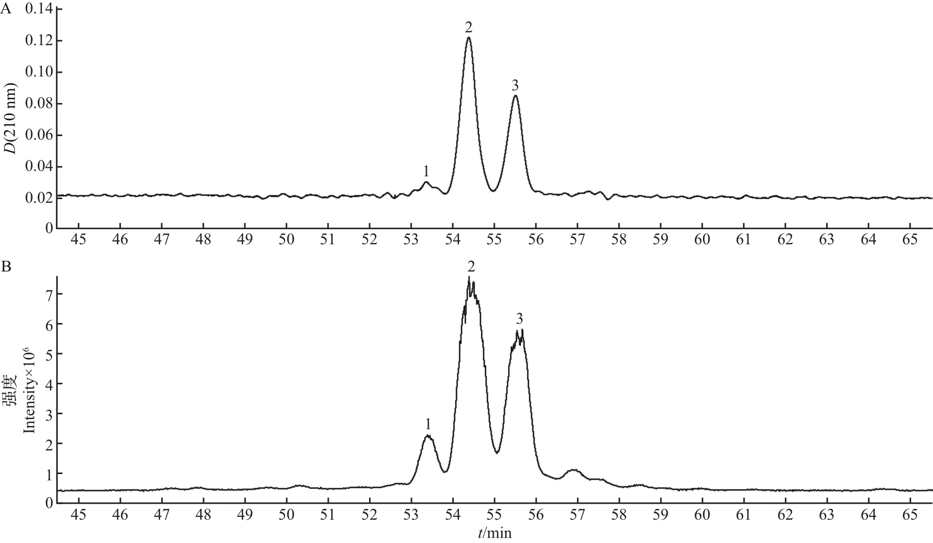
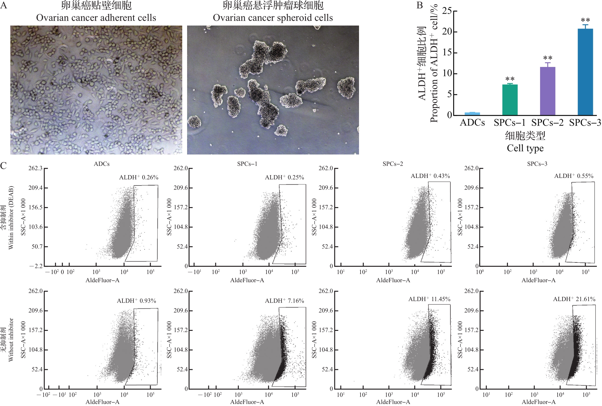
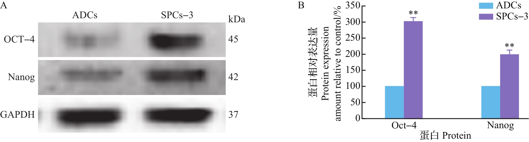
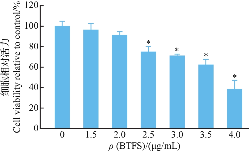
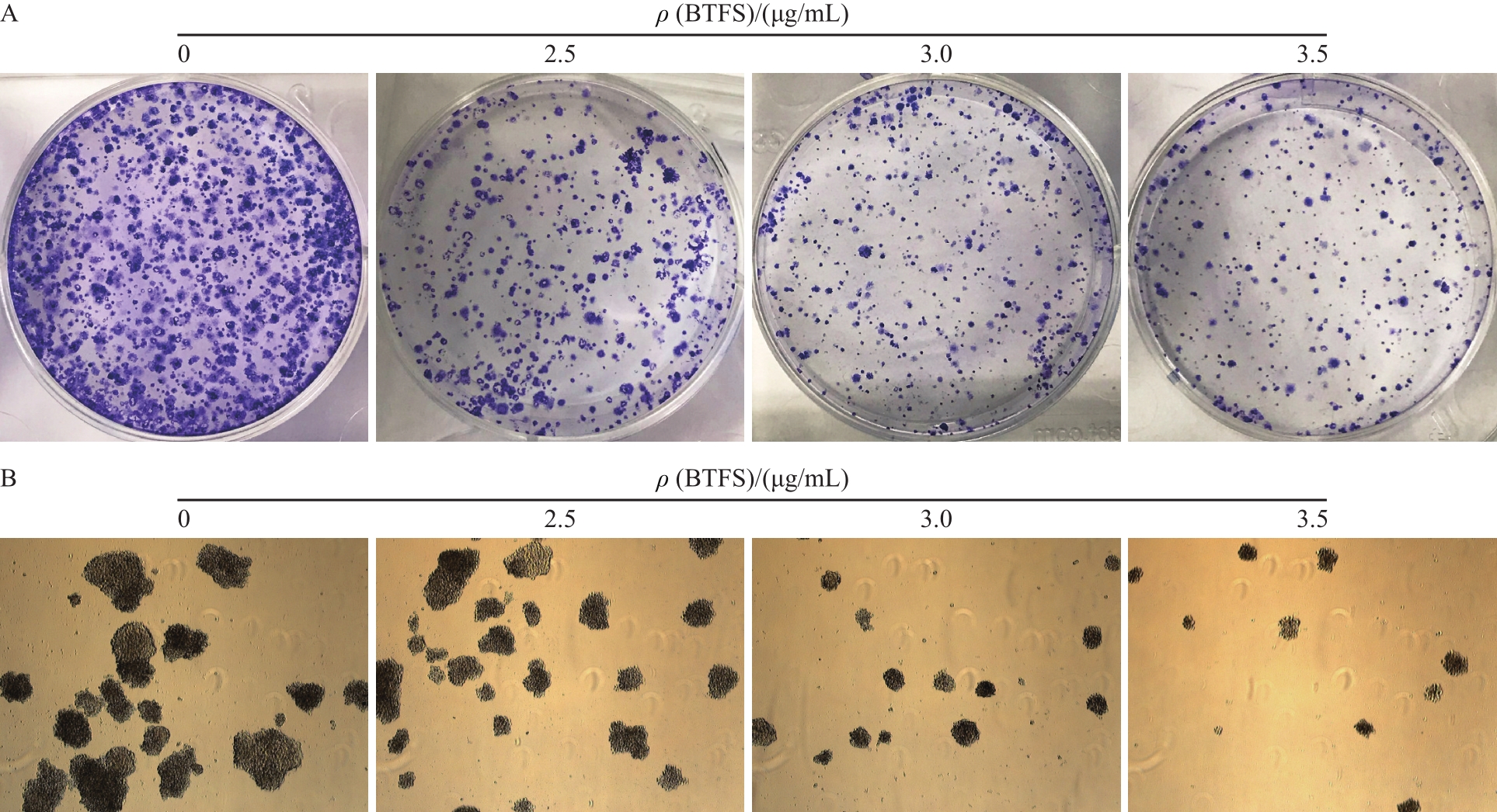
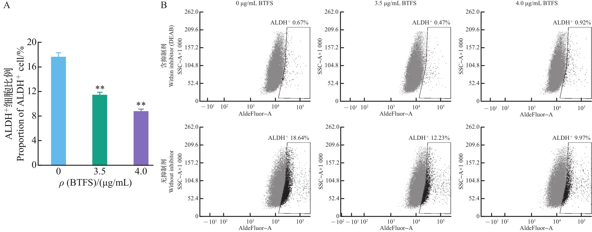
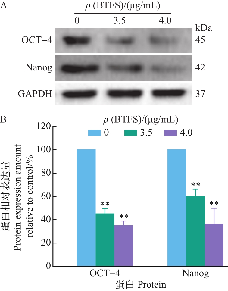
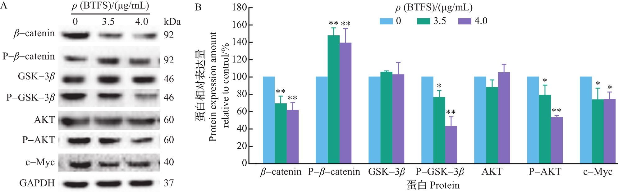
Abstract The effects of tea flower saponin, which was extracted and purified by macroporous resin and preparative liquid chromatography, on the proliferation and traits of human ovarian cancer stem like cells (OCSLCs) and its mechanism were studied. The stem cell marker aldehyde dehydrogenase (ALDH) activity, cell vitality, colony formation capacity, tumor sphere formation capacity and the expression of stemness-related proteins were measured to investigate the effects of tea flower saponin on OCSLCs of A2780/CP70 cells obtained by serum-free suspension culture. Western blot assay was used to investigate the effects of tea flower saponin on Wnt/β-catenin signaling pathway proteins. The results showed that tea flower saponin could decrease the proliferation of OCSLCs by suppressing their cell activity and colony formation ability. Tea flower saponin inhibited the formation of tumor sphere and reduced cell self-renewal ability. The ALDH+ cell proportion and expressions of Oct-4 and Nanog proteins were decreased in OCSLCs treated with tea flower saponin. In addition, tea flower saponin treatment could down-regulate the expression of P-AKT, P-GSK-3β, β-catenin, c-Myc and up-regulate the expression of P-β-catenin to inhibit the Wnt/β-catenin signaling pathway. These results indicate that suppression of the Wnt/β-catenin signaling pathway might be one of the mechanisms by which tea flower saponin inhibits the proliferation of OCSLCs and cancer stem cell traits.
|
|
Received: 02 January 2020
Published: 31 December 2020
|
|
|
|
Corresponding Authors:
Yi Charlie CHEN,Youying TU
E-mail: c.lianfu@foxmail.com;chenyc@ab.edu;youytu@zju.edu.cn
|
茶树花皂苷对卵巢癌干细胞样细胞增殖的影响及机制
利用大孔树脂、制备液相色谱法等从茶树花中提取纯化皂苷,研究茶树花皂苷对人卵巢癌干细胞样细胞(ovarian cancer stem like cells, OCSLCs)增殖生长与干性特征的作用及其机制;采用无血清悬浮培养法获得A2780/CP70细胞的OCSLCs,通过检测干细胞标志物乙醛脱氢酶(aldehyde dehydrogenase, ALDH)活性、细胞活力、细胞克隆和肿瘤球形成能力以及干细胞相关蛋白的表达,分析茶树花皂苷对OCSLCs的作用;同时,采用免疫印迹法分析茶树花皂苷对Wnt/β-联蛋白信号通路蛋白的影响。结果表明:茶树花皂苷可通过降低OCSLCs的细胞活力和克隆形成能力抑制其增殖,抑制肿瘤球形成能力,减弱其自我更新能力;茶树花皂苷可降低OCSLCs中的ALDH+细胞比例,使干性相关蛋白Oct-4和Nanog表达量下降;同时,茶树花皂苷通过下调P-AKT、P-GSK-3β、β-联蛋白、c-Myc蛋白的表达,上调P-β-联蛋白的表达,使Wnt/β-联蛋白信号通路被抑制。综上所述,茶树花皂苷对OCSLCs的增殖和干性具有显著抑制作用,抑制Wnt/β-联蛋白信号通路的活化是其作用机制之一。
关键词:
茶树花,
皂苷,
卵巢癌,
肿瘤干细胞,
Wnt/β-联蛋白信号通路
|
|
| [1] |
MIZUNO T, SUZUKI N, MAKINO H, et al. Cancer stem-like cells of ovarian clear cell carcinoma are enriched in the ALDH-high population associated with an accelerated scavenging system in reactive oxygen species. Gynecologic Oncology, 2015,137(2):299-305. DOI:10.1016/j.ygyno.2014.12.005
doi: 10.1016/j.ygyno.2014
|
|
|
| [2] |
AHMED N, ABUBAKER K, FINDLAY J, et al. Cancerous ovarian stem cells: obscure targets for therapy but relevant to chemoresistance. Journal of Cellular Biochemistry, 2012,114(1):21-34. DOI:10.1002/jcb.24317
doi: 10.1002/jcb.24317
|
|
|
| [3] |
BAPAT S A. Human ovarian cancer stem cells. Reproduction, 2010,140(1):33. DOI:10.1530/REP-09-0389
doi: 10.1530/REP-09-0389
|
|
|
| [4] |
BARBATO L, BOCCHETTI M, BIASE A D, et al. Cancer stem cells and targeting strategies. Cells, 2019,8(8):1-19. DOI:10.3390/cells8080926
doi: 10.3390/cells8080926
|
|
|
| [5] |
BURLESON K M, BOENTE M P, PAMBUCCIAN S E, et al. Disaggregation and invasion of ovarian carcinoma ascites spheroids. Journal of Translational Medicine, 2006,4(1):1-6. DOI:10.1186/1479-5876-4-6
doi: 10.1186/1479-5876-4-6
|
|
|
| [6] |
MUINAO T, PRASANNA H, BORUAH D, et al. Diagnostic and prognostic biomarkers in ovarian cancer and the potential roles of cancer stem cells: an updated review. Experimental Cell Research, 2018,362(1):1-10. DOI:10.1016/j.yexcr.2017.10.018
doi: 10.1016/j.yexcr.2017.10.018
|
|
|
| [7] |
KURODA T, HIROHASHI Y, TORIGOE T, et al. ALDH1-high ovarian cancer stem-like cells can be isolated from serous and clear cell adenocarcinoma cells, and ALDH1 high expression is associated with poor prognosis. PLoS ONE, 2013,8(6):e65158. DOI:10.1371/journal.pone.0065158
doi: 10.1371/journal.pone.0065158
|
|
|
| [8] |
LIU S Y, LIU C F, MIN X Y, et al. Prognostic value of cancer stem cell marker aldehyde dehydrogenase in ovarian cancer: a meta-analysis. PLoS ONE, 2013,8(11):e81050. DOI:10.1371/journal.pone.0081050
doi: 10.1371/journal.pone.0081050
|
|
|
| [9] |
MATSUDA H, NAKAMURA S, MORIKAWA T, et al. New biofunctional effects of the flower buds of Camellia sinensis and its bioactive acylated oleanane-type triterpene oligogly-cosides. Journal of Natural Medicines, 2016,70(4):689-701. DOI:10.1007/s11418-016-1021-1
doi: 10.1007/s11418-016-1021-1
|
|
|
| [10] |
YOSHIKAWA M, MORIKAWA T, YAMAMOTO K, et al. Floratheasaponins A-C, acylated oleanane-type triterpene oligoglycosides with anti-hyperlipidemic activities from flowers of the tea plant (Camellia sinensis). Journal of Natural Products, 2005,68(9):1360-1365. DOI:10.1021/np0580614
doi: 10.1021/np0580614
|
|
|
| [11] |
YOSHIKAWA M, NAKAMURA S, KATO Y, et al. Medicinal flowers. ⅩⅣ. New acylated oleanane-type triterpene oligoglycosides with antiallergic activity from flower buds of Chinese tea plant (Camellia sinensis). Chemical and Pharmaceutical Bulletin, 2007,55(4):598-605. DOI:10.1248/cpb.55.598
doi: 10.1248/cpb.55.598
|
|
|
| [12] |
WANG Y M, REN N, RANKIN G O, et al. Anti-proliferative effect and cell cycle arrest induced by saponins extracted from tea (Camellia sinensis) flower in human ovarian cancer cells. Journal of Functional Foods, 2017,37:310-321. DOI:10.1016/j.jff.2017.08.001
doi: 10.1016/j.jff.2017.08.001
|
|
|
| [13] |
SHEN X, SHI L Z, PAN H B, et al. Identification of triterpenoid saponins in flowers of four Camellia sinensis cultivars from Zhejiang Province: differences between cultivars, developmental stages, and tissues. Industrial Crops and Products, 2017,95:140-147. DOI:10.1016/j.indcrop.2016.10.008
doi: 10.1016/j.indcrop.2016.10
|
|
|
| [14] |
杨丽娟,赖东梅.卵巢癌干细胞及新的治疗策略.现代妇产科进展,2012,21(2):142-144. DOI:10.1002/ijc.31186
YANG L J, LAI D M. Ovarian cancer stem cells and new treatment strategies. Progress in Obstetrics and Gynecology, 2012,21(2):142-144. (in Chinese with English abstract)
doi: 10.1002/ijc.31186
|
|
|
| [15] |
PAN H B, KIM E, RANKIN G O, et al. Theaflavin-3, 3′-digallate inhibits ovarian cancer stem cells via suppressing Wnt/beta-catenin signaling pathway. Journal of Functional Foods, 2018,50:1-7. DOI:10.1016/j.jff.2018.09.021
doi: 10.1016/j.jff.2018.09.021
|
|
|
| [16] |
JIA L Y, XIA H L, CHEN Z D, et al. Anti-proliferation effect of theasaponin E-1 on the ALDH-positive ovarian cancer stem-like cells. Molecules, 2018,23(6):1469-1482. DOI:10.3390/molecules23061469
doi: 10.3390/molecules23061469
|
|
|
| [17] |
SHAN D, YANG X J, LASSUSET H, et al. Distinct expression levels and patterns of stem cell marker, aldehyde dehydrogenase isoform 1 (ALDH1), in human epithelial cancers. PLoS ONE, 2010,5(4):e10277. DOI:10.1371/journal.pone.0010277
doi: 10.1371/journal.pone.0010277
|
|
|
| [18] |
WANG D, LU P, ZHANG H, et al. Oct-4 and NANOG promote the epithelial-mesenchymal transition of breast cancer stem cells and are associated with poor prognosis in breast cancer patients. Oncotarget, 2014,5(21):10803-10815. DOI:10.18632/oncotarget.2506
doi: 10.18632/oncotarget.2506
|
|
|
| [19] |
LEE S, WOTTRICH S, BONAVIDA B. Crosstalks between Raf-kinase inhibitor protein and cancer stem cell transcription factors (Oct4, KLF4, Sox2, Nanog). Tumor Biology, 2017,39(4):1-17. DOI:10.1177/1010428317692253
doi: 10.1177/1010428317692253
|
|
|
| [20] |
KITAGAWA N, MORIKAWA T, MOTAI C, et al. The antiproliferative effect of chakasaponins Ⅰ and Ⅱ, flora-theasaponin A, and epigallocatechin 3-O-gallate isolated from Camellia sinensis on human digestive tract carcinoma cell lines. International Journal of Molecular Sciences, 2016,17(12):1979. DOI:10.3390/ijms17121979
doi: 10.3390/ijms17121979
|
|
|
| [21] |
AREND R C, LONDONO-JOSHI A I, STRAUGHN J M, et al. The Wnt/β-catenin pathway in ovarian cancer: a review. Gynecologic Oncology, 2013,131(3):772-779. DOI:10.1016/j.ygyno2013.09.034
doi: 10.1016/j.ygyno2013.09.034
|
|
|
| [22] |
LYNN R, KAREN D C D. Can stemness and chemoresistance be therapeutically targeted via signaling pathways in ovarian cancer? Cancers, 2018,10(8):241. DOI:10.20944/preprints201806.0262.v1
doi: 10.20944/preprints201806.0262.v1
|
|
|
| [23] |
MACDONALD B T, TAMAI K, HE X. Wnt/β-catenin signaling: components, mechanisms, and diseases. Develop-mental Cell, 2009,17(1):9-26. DOI:10.1016/j.devcel.2009.06.016
doi: 10.1016/j.devcel.2009.06
|
|
|
| [24] |
HE T C, SPARKS A B, RAGO C, et al. Identification of c-MYC as a target of the APC pathway. Science, 1998,281(5382):1509-1512. DOI:10.1126/science.281.5382.1509
doi: 10.1126/science.281.5382.1509
|
|
|
| [25] |
CHEN B J, WU Y L, TANAKA Y, et al. Small molecules targeting c-Myc oncogene: promising anti-cancer therapeutics. International Journal of Biological Sciences, 2014,10(10):1084-1096. DOI:10.7150/ijbs.10190
doi: 10.7150/ijbs.10190
|
|
|
| [26] |
NAIR R, RODEN D L, TEO W S, et al. C-Myc and Her2 cooperate to drive a stem-like phenotype with poor prognosis in breast cancer. Oncogene, 2014,33(30):3992-4002. DOI:10.1038/onc.2013.368
doi: 10.1038/onc.2013.368
|
|
|
| [27] |
SAHLBERG S H, SPIEGELBERG D, GLIMELIUS B, et al. Evaluation of cancer stem cell markers CD133, CD44, CD24: association with AKT isoforms and radiation resistance in colon cancer cells. PLoS ONE, 2014,9(4):e94621. DOI:10.1371/journal.pone.0094621
doi: 10.1371/journal.pone.0094621
|
|
|
| [28] |
NAGARAJ A B, JOSEPH P, KOVALENKO O, et al. Critical role of Wnt/β-catenin signaling in driving epithelial ovarian cancer platinum resistance. Oncotarget, 2015,6(27):23720-23734. DOI:10.18632/oncotarget.4690
doi: 10.18632/oncotarget.4690
|
|
|
| [29] |
ZHANG Y, CHEN S G, WEI C Y, et al. Dietary compound proanthocyanidins from Chinese bayberry (Myrica rubra, Sieb. et Zucc.) leaves inhibit angiogenesis and regulate cell cycle of cisplatin-resistant ovarian cancer cells via targeting Akt pathway. Journal of Functional Foods, 2018,40:573-581. DOI:10.1016/j.jff.2017.11.045
doi: 10.1016/j.jff.2017.11.045
|
|
|
|
Viewed |
|
|
|
Full text
|
|
|
|
|
Abstract
|
|
|
|
|
Cited |
|
|
|
|
| |
Shared |
|
|
|
|
| |
Discussed |
|
|
|
|