| Animal sciences & veterinary medicines |
|

|
|
|
| Mechanism of rapamycin alleviating inflammatory response in bovine mammary epithelial cells |
Lianbin XU( ),Yifei REN(),Wei LAN,Pengfei HOU,Hongyun LIU() ),Yifei REN(),Wei LAN,Pengfei HOU,Hongyun LIU() |
| Institute of Dairy Science, College of Animal Sciences, Zhejiang University, Hangzhou 310058, China |
|
|
|
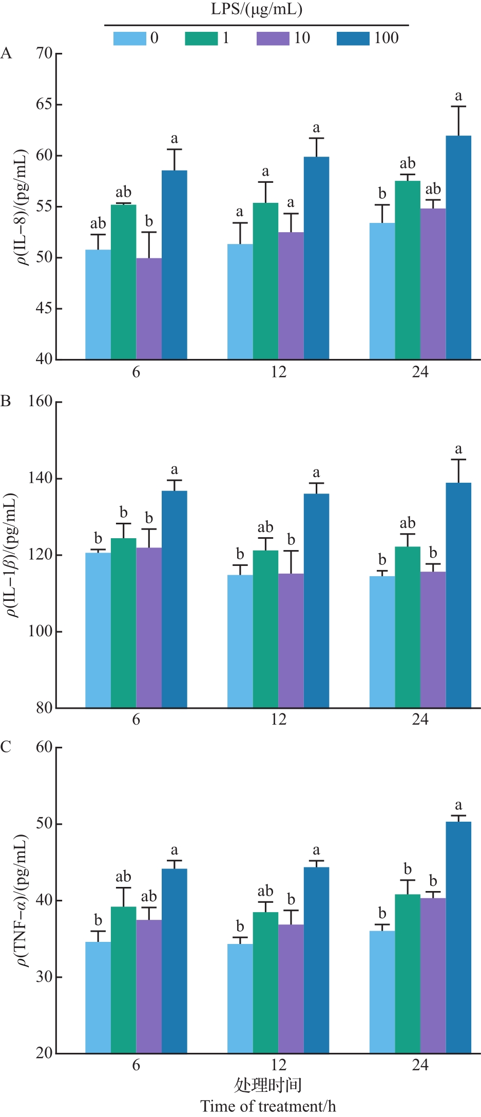
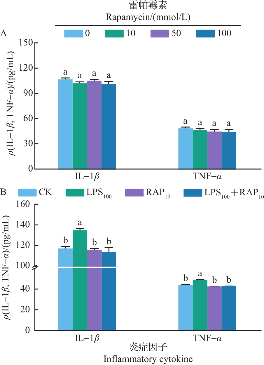
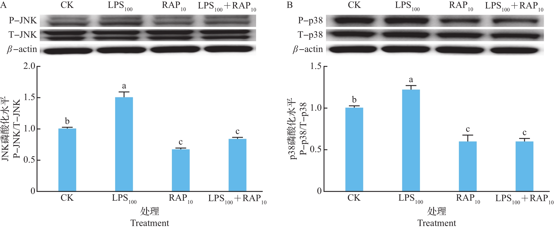
Abstract Mastitis is a frequent disease of dairy cows and results in significant economic losses for dairy producers. In this study, Establishment of inflammation was performed by using different concentrations of lipopolysaccharide (LPS). After that, mammary alveolar cell-T (MAC-T) were randomly cultured in the standard medium (CK), standard medium with 100 μg/mL LPS (LPS100), standard medium with 10 mmol/L rapamycin (RAP10), and standard medium with 100 μg/mL LPS+10 mmol/L RAP (LPS100+RAP10) for 24 h. Cells and culture supernatant were collected at the end of the treatment. The results showed that incubation with LPS for 24 h significantly increased the concentrations of interleukin-8 (IL-8), interleukin-1β (IL-1β) and tumor necrosis factor- α (TNF- α) in MAC-T cells (P<0.05) without affecting the cell viability and apoptosis. Rapamycin addition individually had no significant effects on baseline concentrations of IL-1β and TNF-α (P>0.05), but abolished the increased production of inflammatory cytokines stimulated by LPS (P<0.05). Culturing with LPS increased the phosphorylation and translocation of p65 protein in nuclear factor-κB (NF-κB) signaling pathway, while this elevation was disappeared with the rapamycin supplementation (P<0.05). Phosphorylation levels of c-Jun N-terminal kinase (JNK) and p38 proteins in mitogen-activated protein kinase (MAPK) signaling pathway were lower in LPS100+RAP10 group than that in LPS100 group (P<0.05). These results indicate that rapamycin alleviates LPS-induces inflammatory response in MAC-T cells through the NF-κB/MAPK pathway, which gives a reference for the therapeutic potential of rapamycin in mastitis.
|
|
Received: 29 April 2021
Published: 29 April 2022
|
|
|
|
Corresponding Authors:
Hongyun LIU
E-mail: lianbinxu@zju.edu.cn;hyliu@zju.edu.cn
|
雷帕霉素缓解奶牛乳腺上皮细胞炎症反应的作用机制
乳腺炎是泌乳奶牛的常见疾病,每年给养殖企业造成巨大的经济损失。本研究采用不同浓度的脂多糖(lipopolysaccharide, LPS)诱导奶牛乳腺上皮细胞的炎症反应,之后,在对照培养基(CK)基础上分别添加100 μg/mL LPS(LPS100)、10 mmol/L雷帕霉素(RAP10)及2种物质联合添加(LPS100+RAP10)并处理细胞24 h,结束后收集细胞及上清液用于后续测定。结果表明:在不改变细胞活力和凋亡的情况下,添加100 μg/mL LPS显著提高了上清液中白介素-8(interleukin-8, IL-8)、白介素-1β(interleukin-1β, IL-1β)和肿瘤坏死因子-α(tumor necrosis factor-α, TNF-α)的浓度(P<0.05)。单独添加雷帕霉素不影响奶牛乳腺上皮细胞上清液中炎症因子的浓度(P>0.05),但10 mmol/L雷帕霉素处理显著降低了LPS诱导的IL-1β和TNF-α分泌量(P<0.05)。LPS提高了核因子-κB(nuclear factor-κB, NF-κB)通路中p65蛋白的磷酸化水平,但雷帕霉素的添加消除了这种提高作用(P<0.05)。与LPS100组相比,LPS100+RAP10处理降低了奶牛乳腺上皮细胞丝裂原活化蛋白激酶(mitogen-activated protein kinase, MAPK)通路中c-Jun N末端激酶(c-Jun N-terminal kinase, JNK)和p38蛋白的磷酸化水平(P<0.05)。综上所述,雷帕霉素通过NF-κB/MAPK通路缓解了LPS引起的乳腺上皮细胞炎症反应,该结果可为雷帕霉素用于乳腺炎的治疗提供理论参考。
关键词:
炎症反应,
奶牛乳腺上皮细胞,
核因子-κB/丝裂原活化蛋白激酶,
雷帕霉素
|
|
| [1] |
RUEGG P L. A 100-year review: mastitis detection, management, and prevention[J]. Journal of Dairy Science, 2017, 100(12): 10381-10397. DOI:10.3168/jds.2017-13023
doi: 10.3168/jds.2017-13023
|
|
|
| [2] |
WELLNITZ O, ARNOLD E T, BRUCKMAIER R M. Lipopolysaccharide and lipoteichoic acid induce different immune responses in the bovine mammary gland[J]. Journal of Dairy Science, 2011, 94(11): 5405-5412. DOI:10.3168/jds.2010-3931
doi: 10.3168/jds.2010-3931
|
|
|
| [3] |
KUIPERS A, KOOPS W J, WEMMENHOVE H. Antibiotic use in dairy herds in the Netherlands from 2005 to 2012[J]. Journal of Dairy Science, 2016, 99(2): 1632-1648. DOI:10.3168/jds.2014-8428
doi: 10.3168/jds.2014-8428
|
|
|
| [4] |
MARSHALL B M, LEVY S B. Food animals and antimicrobials: impacts on human health[J]. Clinical Microbiology Reviews, 2011, 24(4): 718-733. DOI:10.1128/CMR.00002-11
doi: 10.1128/CMR.00002-11
|
|
|
| [5] |
OLIVER S P, MURINDA S E. Antimicrobial resistance of mastitis pathogens[J]. Veterinary Clinics of North America: Food Animal Practice, 2012, 28(2): 165-185. DOI:10.1016/j.cvfa.2012.03.005
doi: 10.1016/j.cvfa.2012.03.005
|
|
|
| [6] |
PAKALA R, STABILE E, JANG G J, et al. Rapamycin attenuates atherosclerotic plaque progression in apolipoprotein E knockout mice: inhibitory effect on monocyte chemotaxis[J]. Journal of Cardiovascular Pharmacology, 2005, 46(4): 481-486. DOI:10.1097/01.fjc.0000177985.14305.15
doi: 10.1097/01.fjc.0000177985.14305.15
|
|
|
| [7] |
CHEN W Q, ZHONG L, ZHANG L, et al. Oral rapamycin attenuates inflammation and enhances stability of atherosclerotic plaques in rabbits independent of serum lipid levels[J]. British Journal of Pharmacology, 2009, 156(6): 941-951. DOI:10.1111/j.1476-5381.2008.00102.x
doi: 10.1111/j.1476-5381.2008.00102.x
|
|
|
| [8] |
WARDYN J D, PONSFORD A H, SANDERSON C M. Dissecting molecular cross-talk between Nrf2 and NF-κB response pathways[J]. Biochemical Society Transactions, 2015, 43(4): 621-626. DOI:10.1042/BST20150014
doi: 10.1042/BST20150014
|
|
|
| [9] |
MUNOZ L, AMMIT A J. Targeting p38 MAPK pathway for the treatment of Alzheimer’s disease[J]. Neuropharmacology, 2010, 58(3): 561-568. DOI:10.1016/j.neuropharm.2009.11.010
doi: 10.1016/j.neuropharm.2009.11.010
|
|
|
| [10] |
XU L B, HANIGAN M D, LIN X Y, et al. Effects of jugular infusions of isoleucine, leucine, methionine, threonine, and other amino acids on insulin and glucagon concentrations, mammalian target of rapamycin (mTOR) signaling, and lactational performance in goats[J]. Journal of Dairy Science, 2019, 102(10): 9017-9027. DOI:10.3168/jds.2018-16102
doi: 10.3168/jds.2018-16102
|
|
|
| [11] |
BLUM S E, HELLER E D, LEITNER G. Long term effects of Escherichia coli mastitis[J]. Veterinary Journal, 2014, 201(1): 72-77. DOI:10.1016/j.tvjl.2014.04.008
doi: 10.1016/j.tvjl.2014.04.008
|
|
|
| [12] |
VALLABHAPURAPU S, KARIN M. Regulation and function of NF-κB transcription factors in the immune system[J]. Annual Review of Immunology, 2009, 27(1): 693-733. DOI:10.1146/annurev.immunol.021908.132641
doi: 10.1146/annurev.immunol.021908.132641
|
|
|
| [13] |
SOKOLOVA O, NAUMANN M. NF-κB signaling in gastric cancer[J]. Toxins (Basel), 2017, 9(4): 119. DOI:10.3390/toxins9040119
doi: 10.3390/toxins9040119
|
|
|
| [14] |
CHEN J Y, STARK L A. Crosstalk between NF-κB and nucleoli in the regulation of cellular homeostasis[J]. Cells, 2018, 7(10): 157. DOI:10.3390/cells7100157
doi: 10.3390/cells7100157
|
|
|
| [15] |
BAO W L, WANG Y F, FU Y T, et al. mTORC1 regulates flagellin-induced inflammatory response in macrophages[J]. PLoS ONE, 2015, 10(5): e0125910. DOI:10.1371/journal.pone.0125910
doi: 10.1371/journal.pone.0125910
|
|
|
| [16] |
DITTRICH E, SCHMALDIENST S, SOLEIMAN A, et al. Rapamycin-associated post-transplantation glomerulonephritis and its remission after reintroduction of calcineurin-inhibitor therapy[J]. Transplant International, 2004, 17(4): 215-220. DOI:10.1007/s00147-004-0700-0
doi: 10.1007/s00147-004-0700-0
|
|
|
| [17] |
THAUNAT O, BEAUMONT C, CHATENOUD L, et al. Anemia after late introduction of sirolimus may correlate with biochemical evidence of a chronic inflammatory state[J]. Transplantation, 2005, 80(9): 1212-1219. DOI:10.1097/01.tp.0000179106.07382.6a
doi: 10.1097/01.tp.0000179106.07382.6a
|
|
|
| [18] |
BRAICU C, BUSE M, BUSUIOC C, et al. A comprehensive review on MAPK: a promising therapeutic target in cancer[J]. Cancers (Basel), 2019, 11(10): 1618. DOI:10.3390/cancers11101618
doi: 10.3390/cancers11101618
|
|
|
| [19] |
BACHSTETTER A D, XING B, DE ALMEIDA L, et al. Microglial p38α MAPK is a key regulator of proinflammatory cytokine up-regulation induced by toll-like receptor (TLR) ligands or beta-amyloid (Aβ)[J]. Journal of Neuroinflammation, 2011, 8: 79. DOI:10.1186/1742-2094-8-79
doi: 10.1186/1742-2094-8-79
|
|
|
|
Viewed |
|
|
|
Full text
|
|
|
|
|
Abstract
|
|
|
|
|
Cited |
|
|
|
|
| |
Shared |
|
|
|
|
| |
Discussed |
|
|
|
|