| Article |
|


|
|
|
| Adenovirus-mediated GDF-5 promotes the extracellular matrix expression in degenerative nucleus pulposus cells |
Xu-wei Luo,Kang Liu,Zhu Chen,Ming Zhao,Xiao-wei Han,Yi-guang Bai,Gang Feng( ) ) |
| Research Institute of Tissue Engineering and Stem Cells, Nanchong Central Hospital and the Second Clinical Institute of North Sichuan Medical College, Nanchong 637000, China |
|
|
|
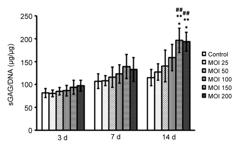
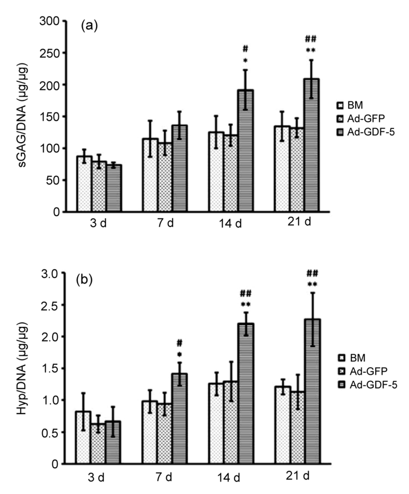
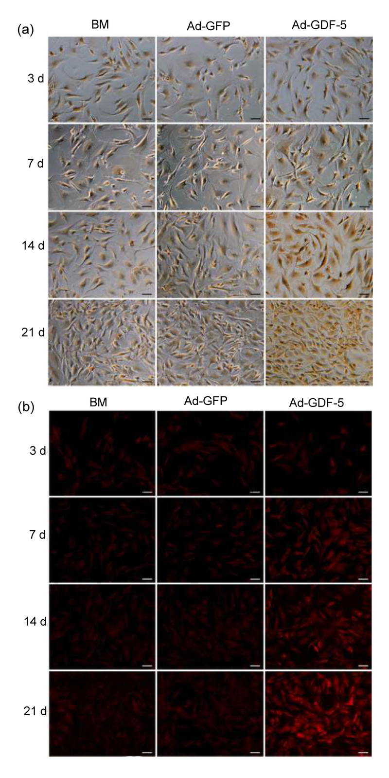
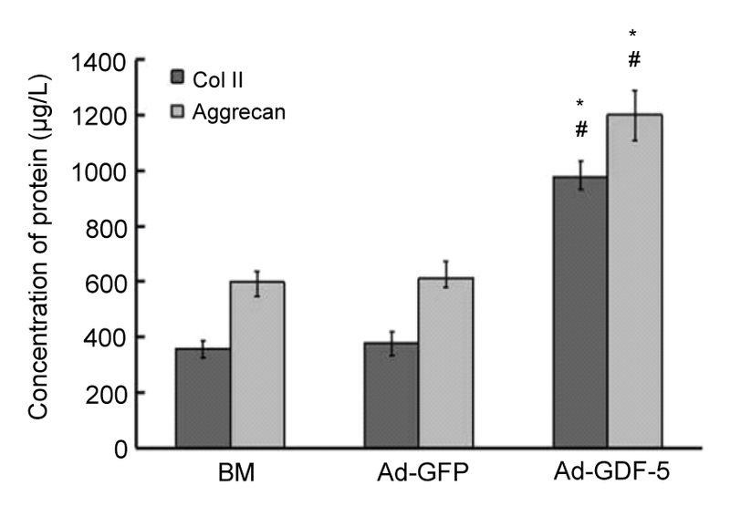
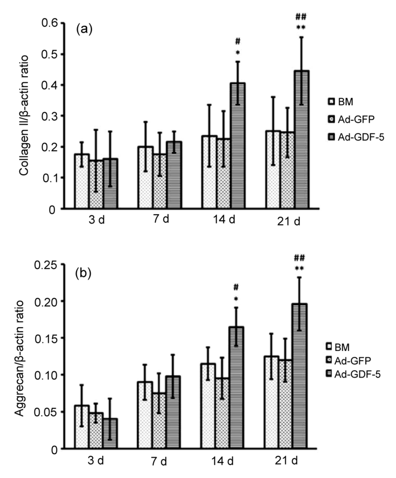
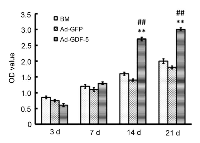
Abstract Objective: To construct a recombinant adenovirus vector-carrying human growth and differentiation factor-5 (GDF-5) gene, investigate the biological effects of adenovirus-mediated GDF-5 (Ad-GDF-5) on extracellular matrix (ECM) expression in human degenerative disc nucleus pulposus (NP) cells, and explore a candidate gene therapy method for intervertebral disc degeneration (IDD). Methods: Human NP cells of a degenerative disc were isolated, cultured, and infected with Ad-GDF-5 using the AdEasy-1 adenovirus vector system. On Days 3, 7, 14, and 21, the contents of the sulfated glycosaminoglycan (sGAG), deoxyribonucleic acid (DNA) and hydroxyproline (Hyp), synthesis of proteoglycan and collagen II, gene expression of collagen II and aggrecan, and NP cell proliferation were assessed. Results: The adenovirus was an effective vehicle for gene delivery with prolonged expression of GDF-5. Biochemical analysis revealed increased sGAG and Hyp contents in human NP cells infected by Ad-GDF-5 whereas there was no conspicuous change in basal medium (BM) or Ad-green fluorescent protein (GFP) groups. Only cells in the Ad-GDF-5 group promoted the production of ECM, as demonstrated by the secretion of proteoglycan and up-regulation of collagen II and aggrecan at both protein and mRNA levels. The NP cell proliferation was significantly promoted. Conclusions: The data suggest that Ad-GDF-5 gene therapy is a potential treatment for IDD, which restores the functions of degenerative intervertebral disc through enhancing the ECM production of human NP cells.
|
|
Received: 30 July 2015
Published: 01 January 2016
|
| Fund: Project supported by the National Natural Science Foundation of China(Nos. 81171472, 81201407);the Innovation Team Project of Sichuan Provincial Education Department(No. 13TD0030);the Major Transformation Cultivation Project of Sichuan Provincial Education Department(No. 15CZ0021);the Science and Technology Project of Nanchong City, China(No. 14A0021) |
|
Corresponding Authors:
Gang Feng
E-mail: fenggangncch@163.com
|
|
|
| [1] |
Bucher C, Gazdhar A, Benneker LM. Nonviral gene delivery of growth and differentiation factor 5 to human mesenchymal stem cells injected into a 3D bovine intervertebral disc organ culture system. Stem Cells Int. 2013, 2013:326828 (Available from: http://dx.doi.org/10.1155/2013/326828)
doi: 10.1155/2013/326828
pmid: 3885261
|
|
|
| [2] |
Chubinskaya S, Hurtig M, Rueger DC. OP-1/BMP-7 in cartilage repair. Int Orthop. 2007, 31(6):773-781. (Available from: http://dx.doi.org/10.1007/s00264-007-0423-9)
doi: 10.1007/s00264-007-0423-9
|
|
|
| [3] |
Costello DJ, O'Keeffe GW, Hurley FM. Transplantation of novel human GDF5-expressing CHO cells is neuroprotective in models of Parkinson’s disease. J Cell Mol Med. 2012, 16(10):2451-2460. (Available from: http://dx.doi.org/10.1111/j.1582-4934.2012.01562.x)
doi: 10.1111/j.1582-4934.2012.01562.x
pmid: 22436046
|
|
|
| [4] |
Cui M, Wan Y, Anderson DG. Mouse growth and differentiation factor-5 protein and DNA therapy potentiates intervertebral disc cell aggregation and chondrogenic gene expression. Spine J. 2008, 8(2):287-295. (Available from: http://dx.doi.org/10.1016/j.spinee.2007.05.012)
doi: 10.1016/j.spinee.2007.05.012
|
|
|
| [5] |
Daans M, Luyten FP, Lories RJ. GDF5 deficiency in mice is associated with instability-driven joint damage, gait and subchondral bone changes. Ann Rheum Dis. 2011, 70(1):208-213. (Available from: http://dx.doi.org/10.1136/ard.2010.134619)
doi: 10.1136/ard.2010.134619
|
|
|
| [6] |
Elkasrawy MN, Hamrick MW. Myostatin (GDF-8) as a key factor linking muscle mass and bone structure. J Musculoskelet Neuronal Interact. 2010, 10(1):56-63
|
|
|
| [7] |
Ellman MB, An HS, Muddasani P. Biological impact of the fibroblast growth factor family on articular cartilage and intervertebral disc homeostasis. Gene. 2008, 420(1):82-89. (Available from: http://dx.doi.org/10.1016/j.gene.2008.04.019)
doi: 10.1016/j.gene.2008.04.019
pmid: 2525607
|
|
|
| [8] |
Eskola PJ, Lemmel? S, Kjaer P. Genetic association studies in lumbar disc degeneration: a systematic review. PLoS ONE. 2012, 7(11):e49995 (Available from: http://dx.doi.org/10.1371/journal.pone.0049995)
doi: 10.1371/journal.pone.0049995
pmid: 3503778
|
|
|
| [9] |
Farndale RW, Buttle DJ, Barrett AJ. Improved quantitation and discrimination of sulphated glycosaminoglycans by use of dimethylmethylene blue. Biochim Biophys Acta. 1986, 883(2):173-177. (Available from: http://dx.doi.org/10.1016/0304-4165(86)90306-5)
doi: 10.1016/0304-4165(86)90306-5
|
|
|
| [10] |
Feng G, Wan Y, Balian G. Adenovirus-mediated expression of growth and differentiation factor-5 promotes chondrogenesis of adipose stem cells. Growth Factors. 2008, 26(3):132-142. (Available from: http://dx.doi.org/10.1080/08977190802105917)
doi: 10.1080/08977190802105917
pmid: 18569021
|
|
|
| [11] |
Feng G, Wan Y, Shen FH. Nucleus pulposus explant culture model. J Orthop Res. 2009, 27(6):814-819. (Available from: http://dx.doi.org/10.1002/jor.20803)
doi: 10.1002/jor.20803
pmid: 19030173
|
|
|
| [12] |
Freemont AJ. The cellular pathobiology of the degenerate intervertebral disc and discogenic back pain. Rheumatology. 2009, 48(1):5-10. (Available from: http://dx.doi.org/10.1093/rheumatology/ken396)
doi: 10.1093/rheumatology/ken396
pmid: 18854342
|
|
|
| [13] |
Hanley ENJr, Herkowitz HN, Kirkpatrick JS. Debating the value of spine surgery. J Bone Joint Surg Am. 2010, 92(5):1293-1304. (Available from: http://dx.doi.org/10.2106/JBJS.I.01439)
doi: 10.2106/JBJS.I.01439
pmid: 20439681
|
|
|
| [14] |
Hogan M, Girish K, James R. Growth differentiation factor-5 regulation of extracellular matrix gene expression in murine tendon fibroblasts. J Tissue Eng Regen Med. 2011, 5(3):191-200. (Available from: http://dx.doi.org/10.1002/term.304)
doi: 10.1002/term.304
pmid: 20653042
|
|
|
| [15] |
Hua G, Haiping Z, Baorong H. Effect of ulinastatin on the expression of iNOS, MMP-2, and MMP-3 in degenerated nucleus pulposus cells of rabbits. Connect Tissue Res. 2013, 54(1):29-33. (Available from: http://dx.doi.org/10.3109/03008207.2012.716882)
doi: 10.3109/03008207.2012.716882
|
|
|
| [16] |
Kaneyama S, Nishida K, Takada T. Fas ligand expression on human nucleus pulposus cells decreases with disc degeneration processes. J Orthop Sci. 2008, 13(2):130-135. (Available from: http://dx.doi.org/10.1007/s00776-007-1204-4)
doi: 10.1007/s00776-007-1204-4
|
|
|
| [17] |
Kim JS, Ellman MB, An HS. Lactoferricin mediates anabolic and anti-catabolic effects in the intervertebral disc. J Cell Physiol. 2012, 227(4):1512-1520. (Available from: http://dx.doi.org/10.1002/jcp.22867)
doi: 10.1002/jcp.22867
|
|
|
| [18] |
Kim YJ, Sah RL, Doong JY. Fluorometric assay of DNA in cartilage explants using Hoechst 33258. Anal Biochem. 1988, 174(1):168-176. (Available from: http://dx.doi.org/10.1016/0003-2697(88)90532-5)
doi: 10.1016/0003-2697(88)90532-5
pmid: 2464289
|
|
|
| [19] |
Larson III JW, Levicoff EA, Gilbertson LG. Biologic modification of animal models of intervertebral disc degeneration. J Bone Joint Surg Am. 2006, 88(Suppl. 2):83-88. (Available from: http://dx.doi.org/10.2106/JBJS.F.00043)
doi: 10.2106/JBJS.F.00043
pmid: 16595450
|
|
|
| [20] |
Li X, Leo BM, Beck G. Collagen and proteoglycan abnormalities in the GDF-5-deficient mice and molecular changes when treating disk cells with recombinant growth factor. Spine. 2004, 29(20):2229-2234. (Available from: http://dx.doi.org/10.1097/01.brs.0000142427.82605.fb)
doi: 10.1097/01.brs.0000142427.82605.fb
pmid: 15480133
|
|
|
| [21] |
Liang H, Ma SY, Feng G. Therapeutic effects of adenovirus-mediated growth and differentiation factor-5 in a mice disc degeneration model induced by annulus needle puncture. Spine J. 2010, 10(1):32-41. (Available from: http://dx.doi.org/10.1016/j.spinee.2009.10.006)
doi: 10.1016/j.spinee.2009.10.006
|
|
|
| [22] |
Masuda K. Biological repair of the degenerated intervertebral disc by the injection of growth factors. Eur Spine J. 2008, 17(Suppl. 4):441-451. (Available from: http://dx.doi.org/10.1007/s00586-008-0749-z)
doi: 10.1007/s00586-008-0749-z
pmid: 2587664
|
|
|
| [23] |
Melrose J, Shu C, Young C. Mechanical destabilization induced by controlled annular incision of the intervertebral disc dysregulates metalloproteinase expression and induces disc degeneration. Spine. 2012, 37(1):18-25. (Available from: http://dx.doi.org/10.1097/BRS.0b013e31820cd8d5)
doi: 10.1097/BRS.0b013e31820cd8d5
pmid: 22179320
|
|
|
| [24] |
Moore YR, Dickinson DP, Wikesj? UM. Growth/differentiation factor-5: a candidate therapeutic agent for periodontal regeneration A review of pre-clinical data. J Clin Periodontol. 2010, 37(3):288-298. (Available from: http://dx.doi.org/10.1111/j.1600-051X.2009.01527.x)
doi: 10.1111/j.1600-051X.2009.01527.x
pmid: 20088982
|
|
|
| [25] |
Nishida K, Kang JD, Suh JK. Adenovirus-mediated gene transfer to nucleus pulposus cells. Implications for the treatment of intervertebral disc degeneration. Spine. 1998, 23(22):2437-2442. (Available from: http://dx.doi.org/10.1097/00007632-199811150-00016)
doi: 10.1097/00007632-199811150-00016
pmid: 9836359
|
|
|
| [26] |
Nishida K, Kang JD, Gilbertson LG. Modulation of the biologic activity of the rabbit intervertebral disc by gene therapy: an in vivo study of adenovirus-mediated transfer of the human transforming growth factor β1 encoding gene. Spine. 1999, 24(23):2419-2425. (Available from: http://dx.doi.org/10.1097/00007632-199912010-00002)
doi: 10.1097/00007632-199912010-00002
pmid: 10626303
|
|
|
| [27] |
Pfirrmann CW, Metzdorf A, Zanetti M. Magnetic resonance classification of lumbar intervertebral disc degeneration. Spine. 2001, 26(17):1873-1878. (Available from: http://dx.doi.org/10.1097/00007632-200109010-00011)
doi: 10.1097/00007632-200109010-00011
pmid: 11568697
|
|
|
| [28] |
Phillips FM, Slosar PJ, Youssef JA. Lumbar spine fusion for chronic low back pain due to degenerative disc disease: a systematic review. Spine. 2013, 38(7):E409-E422. (Available from: http://dx.doi.org/10.1097/BRS.0b013e3182877f11)
doi: 10.1097/BRS.0b013e3182877f11
pmid: 23334400
|
|
|
| [29] |
Saiga K, Furumatsu T, Yoshida A. Combined use of bFGF and GDF-5 enhances the healing of medial collateral ligament injury. Biochem Biophys Res Commun. 2010, 402(2):329-334. (Available from: http://dx.doi.org/10.1016/j.bbrc.2010.10.026)
doi: 10.1016/j.bbrc.2010.10.026
pmid: 20937261
|
|
|
| [30] |
Shimer AL, Chadderdon RC, Gilbertson LG. Gene therapy approaches for intervertebral disc degeneration. Spine. 2004, 29(23):2770-2778. (Available from: http://dx.doi.org/10.1097/01.brs.0000146455.11253.08)
doi: 10.1097/01.brs.0000146455.11253.08
|
|
|
| [31] |
Smith LJ, Nerurkar NL, Choi KS. Degeneration and regeneration of the intervertebral disc: lessons from development. Dis Model Mech. 2011, 4(1):31-41. (Available from: http://dx.doi.org/10.1242/dmm.006403)
doi: 10.1242/dmm.006403
|
|
|
| [32] |
Sobajima S, Kim JS, Gilbertson LG. Gene therapy for degenerative disc disease. Gene Ther. 2004, 11(4):390-401. (Available from: http://dx.doi.org/10.1038/sj.gt.3302200)
doi: 10.1038/sj.gt.3302200
pmid: 14724681
|
|
|
| [33] |
Stoyanov JV, Gantenbein-Ritter B, Bertolo A. Role of hypoxia and growth and differentiation factor-5 on differentiation of human mesenchymal stem cells towards intervertebral nucleus pulposus-like cells. Eur Cell Mater. 2011, 21 533-547
doi: 10.22203/eCM.v021a40
pmid: 21710444
|
|
|
| [34] |
Walsh AJ, Bradford DS, Lotz JC. In vivo growth factor treatment of degenerated intervertebral discs. Spine. 2004, 29(2):156-163. (Available from: http://dx.doi.org/10.1097/01.BRS.0000107231.67854.9F)
doi: 10.1097/01.BRS.0000107231.67854.9F
pmid: 14722406
|
|
|
| [35] |
Wang H, Kroeber M, Hanke M. Release of active and depot GDF-5 after adenovirus-mediated overexpression stimulates rabbit and human intervertebral disc cells. J Mol Med. 2004, 82(2):126-134. (Available from: http://dx.doi.org/10.1007/s00109-003-0507-y)
doi: 10.1007/s00109-003-0507-y
|
|
|
| [36] |
Williams FM, Popham M, Hart DJ. GDF5 single-nucleotide polymorphism rs143383 is associated with lumbar disc degeneration in Northern European women. Arthritis Rheum. 2011, 63(3):708-712. (Available from: http://dx.doi.org/10.1002/art.30169)
doi: 10.1002/art.30169
pmid: 3498734
|
|
|
| [37] |
Zaidi SH, Huang Q, Momen A. Growth differentiation factor 5 regulates cardiac repair after myocardial infarction. J Am Coll Cardiol. 2010, 55(2):135-143. (Available from: http://dx.doi.org/10.1016/j.jacc.2009.08.041)
doi: 10.1016/j.jacc.2009.08.041
pmid: 20117381
|
|
|
|
Viewed |
|
|
|
Full text
|
|
|
|
|
Abstract
|
|
|
|
|
Cited |
|
|
|
|
| |
Shared |
|
|
|
|
| |
Discussed |
|
|
|
|