| Special Topic: Crop Phenotyping Technologies and Applications |
|

|
|
|
| Diagnosis of citrus leaf canker disease based on naive Bayesian classification |
Meiyan SHU1,Jiaxi WEI1,2,3,Yeying ZHOU1,Qizhou DONG1,Haochong CHEN1,Zhigang HUANG2( ),Yuntao MA1 ),Yuntao MA1 |
1.College of Land Science and Technology, China Agricultural University, Beijing 100193, China
2.College of Agriculture, Guangxi University, Nanning 530004, China
3.Beijing Municipal Veterans Affairs Bureau, Beijing 100020, China |
|
|
|
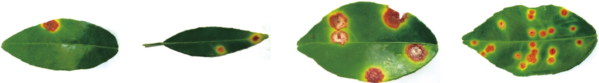
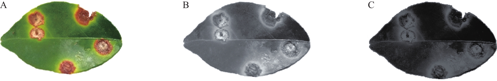
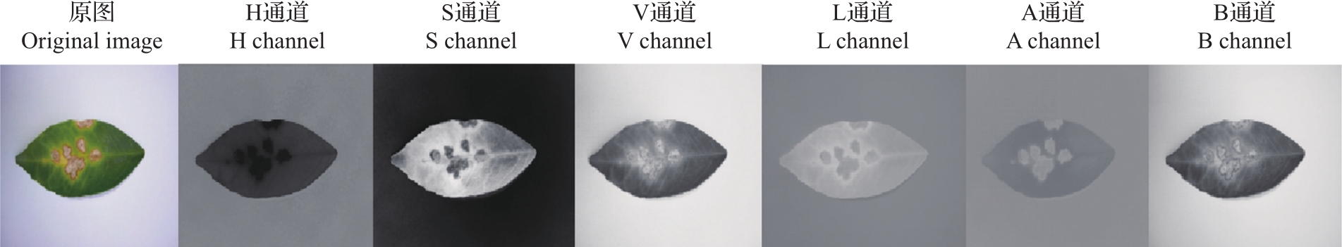
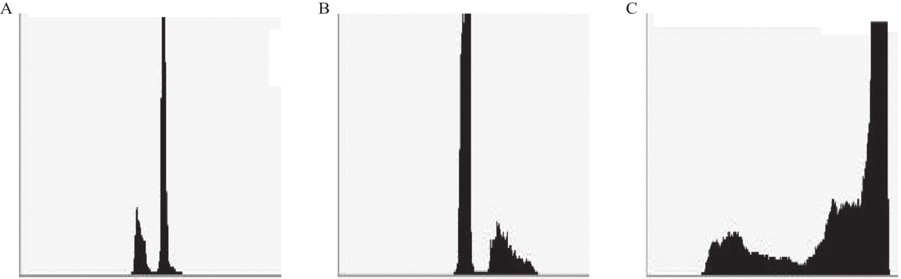
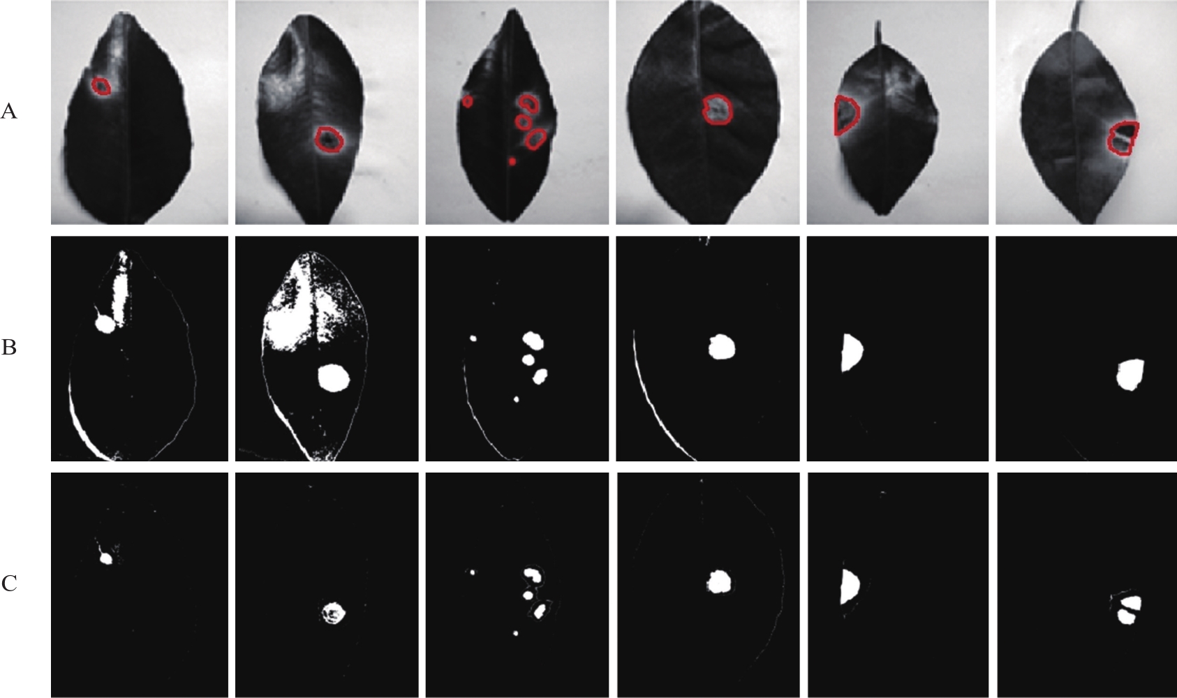
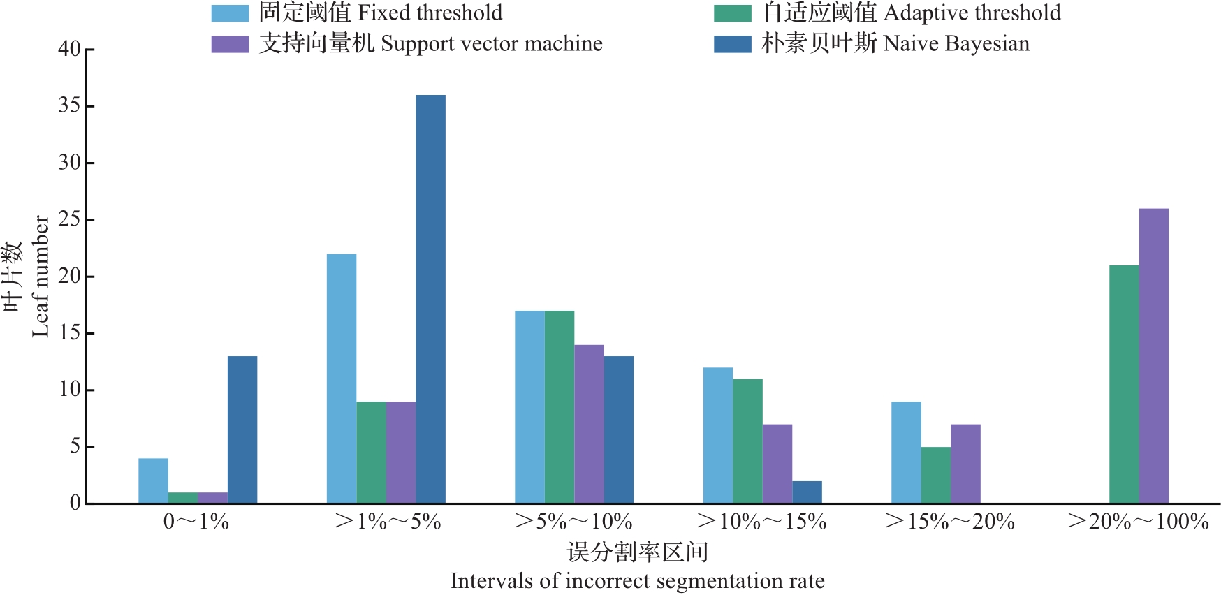
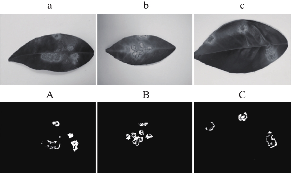
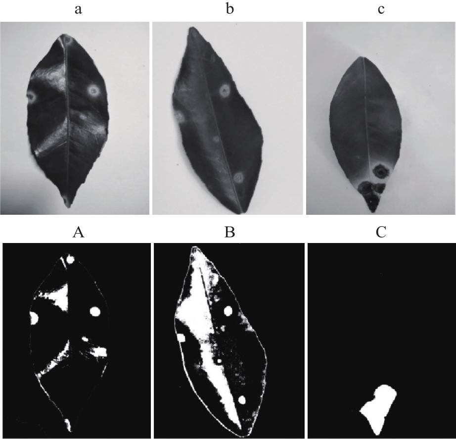
Abstract In order to recognize citrus leaf canker disease accurately and quickly, a diagnosis method of citrus leaf canker disease based on naive Bayesian classification was proposed. The digital images of leaves with different severities of citrus leaf canker disease were used as the data source. According to the characteristics of color space, a disease spot recognition model based on naive Bayesian classification was established for rapid diagnosis of citrus leaf canker disease, and the diagnostic abilities of naive Bayesian classification, fixed threshold, adaptive threshold and support vector machine for citrus leaf canker disease were compared. The results showed that the method based on naive Bayesian classification was effective in the segmentation of citrus leaf canker disease, and the incorrect segmentation rate was only 3.58%, which was far better than the threshold methods and support vector machine. In terms of performance efficiency, the time order of the four algorithms was fixed threshold method<adaptive threshold<naive Bayesian<support vector machine, all of which were within a reasonable range. Combined with the preparation time, naive Bayesian method had the best performance efficiency. Therefore, the naive Bayesian classification algorithm has a rapid and accurate application ability in the diagnosis of citrus leaf canker disease, and can provide a new way for the accurate diagnosis of fruit tree disease severities.
|
|
Received: 01 April 2021
Published: 02 September 2021
|
|
|
|
Corresponding Authors:
Zhigang HUANG
E-mail: hzg@gxu.edu.cn
|
基于朴素贝叶斯分类的柑橘叶片溃疡病诊断
为实现准确、快速地识别柑橘叶片溃疡病,提出一种基于朴素贝叶斯分类的柑橘叶片溃疡病诊断方法。基于不同病害程度的叶片数码图像,根据颜色空间特征,构建基于朴素贝叶斯的柑橘叶片溃疡病斑识别模型,并对比分析朴素贝叶斯分类、固定阈值分割、自适应阈值分割、支持向量机分割对柑橘叶片溃疡病的诊断能力。结果表明:基于朴素贝叶斯分类的柑橘叶片溃疡病斑分割效果较好,误分割率仅为3.58%,远远优于阈值法和支持向量机。在运行效率方面,4种算法耗时排序为固定阈值法<自适应阈值法<朴素贝叶斯法<支持向量机法,但均在较合理的范围内;结合前期准备时间,朴素贝叶斯法的运行效率最佳。综上所述,朴素贝叶斯分类算法在柑橘叶片溃疡病诊断方面具有快速、精准的应用能力,可以为果树从业者精确诊断果树病害严重度提供新思路。
关键词:
柑橘,
溃疡病,
朴素贝叶斯分类,
阈值分割
|
|
| [1] |
苏坚任.柑橘溃疡病的综合防治分析与研究.农业与技术,2018,38(1):85-86.
SU J R. Analysis and study on comprehensive control of citrus canker disease. Agriculture and Technology, 2018,38(1):85-86. (in Chinese)
|
|
|
| [2] |
RAMOS A P, TALHINHAS P, SREENIVASAPRASAD S, et al. Characterization of Colletotrichum gloeosporioides, as the main causal agent of citrus anthracnose, and C. karstii as species preferentially associated with lemon twig dieback in Portugal. Phytoparasitica, 2016,44(4):549-561. DOI:10.1007/s12600-016-0537-y
doi: 10.1007/s12600-016-0537-y
|
|
|
| [3] |
WENG H Y, Lü J W, CEN H Y, et al. Hyperspectral reflectance imaging combined with carbohydrate metabolism analysis for diagnosis of citrus Huanglongbing in different seasons and cultivars. Sensors and Actuators B: Chemical, 2018,275(1):50-60. DOI:10.1016/j.snb.2018.08.020
doi: 10.1016/j.snb.2018.08.020
|
|
|
| [4] |
SHARIF M, KHAN M A, LQBAL Z, et al. Detection and classification of citrus diseases in agriculture based on optimized weighted segmentation and feature selection. Computers and Electronics in Agriculture, 2018,150:220-234. DOI:10.1016/j.compag.2018.04.023
doi: 10.1016/j.compag.2018.04.023
|
|
|
| [5] |
QIN J W, BURKS T F, RITENOUR M A, et al. Detection of citrus canker using hyperspectral reflectance imaging with spectral information divergence. Journal of Food Engineering, 2009,93(2):183-191. DOI:10.1016/j.jfoodeng.2009.01.014
doi: 10.1016/j.jfoodeng.2009.01.014
|
|
|
| [6] |
DUAN S, JIA H G, PANG Z Q, et al. Functional characterization of the citrus canker susceptibility gene CsLOB1. Molecular Plant Pathology, 2018,19(8):1908-1916. DOI:10.1111/mpp.12667
doi: 10.1111/mpp.12667
|
|
|
| [7] |
ABDULRIDHA J, BATUMAN O, AMPATZIDIS Y. UAV-based remote sensing technique to detect citrus canker disease utilizing hyperspectral imaging and machine learning. Remote Sensing, 2019,11(11):1373. DOI:10.3390/rs11111373
doi: 10.3390/rs11111373
|
|
|
| [8] |
赵云,宋寅卯,刁智华.基于图像技术的农作物病害识别.河南农业,2013(8):62-64.
ZHAO Y, SONG Y M, DIAO Z H. Identification of agricultural plant diseases based on image technology. Agriculture of Henan, 2013(8):62-64. (in Chinese)
|
|
|
| [9] |
GOWEN A A, O’DONNELL C P, CULLEN P J, et al. Hyperspectral imaging: an emerging process analytical tool for food quality and safety control. Trends in Food Science and Technology, 2007,18(12):590-598. DOI:10.1016/j.tifs.2007.06.001
doi: 10.1016/j.tifs.2007.06.001
|
|
|
| [10] |
ALEIXOS N, BLASCO J, NAVARRON F, et al. Multispectral inspection of citrus in real-time using machine vision and digital signal processors. Computers and Electronics in Agriculture, 2002,33(2):121-137. DOI:10.1016/S0168-1699(02)00002-9
doi: 10.1016/S0168-1699(02)00002-9
|
|
|
| [11] |
张明,王生荣,郭小燕.基于混合蛙跳算法的马铃薯病害图像分割优化.植物保护学报,2018,45(3):478-488. DOI:10.13802/j.cnki.zwbhxb.2018.2017111
ZHANG M, WANG S R, GUO X Y. Optimization of potato disease image segmentation based on hybrid leapfrog algorithm. Journal of Plant Protection, 2018,45(3):478-488. (in Chinese with English abstract)
doi: 10.13802/j.cnki.zwbhxb.2018.2017111
|
|
|
| [12] |
KUMAR A, LEE W S, EHSANI R J, et al. Citrus greening disease detection using aerial hyperspectral and multispectral imaging techniques. Journal of Applied Remote Sensing, 2012,6(1):063542. DOI:10.1117/1.JRS.6.063542
doi: 10.1117/1.JRS.6.063542
|
|
|
| [13] |
KIM D G, BURKS T F, QIN J W, et al. Classification of grapefruit peel diseases using color texture feature analysis. International Journal of Agricultural and Biological Engineering, 2009,2(3):41-50. DOI:10.13031/2013.24555
doi: 10.13031/2013.24555
|
|
|
| [14] |
PANMANAS S, YUKI H, MUNEHIRO T. Study on nondestructive evaluation methods for defect pods for green soybean processing by near-infrared spectroscopy. Journal of Food Engineering, 2009,93(4):502-512. DOI:10.1016/j.jfooding.2009.02.019
doi: 10.1016/j.jfooding.2009.02.019
|
|
|
| [15] |
WANG X, ZHANG M, ZHU J, et al. Spectral prediction of Phytophthora infestans infection on tomatoes using artificial neural network (ANN). International Journal of Remote Sensing, 2008,29(6):1693-1706. DOI:10.1080/01431160701281007
doi: 10.1080/01431160701281007
|
|
|
| [16] |
LUO J H, HUANG W J, ZHAO J L, et al. Detecting aphid density of winter wheat leaf using hyperspectral measure-ments. IEEE Journal of Selected Topics in Applied Earth Observations and Remote Sensing, 2013,6(2):690-698. DOI:10.1109/JSTARS.2013.2248345
doi: 10.1109/JSTARS.2013.2248345
|
|
|
| [17] |
ASHOURLOO D, AGHIGHI H, MATKAN A A, et al. An investigation into machine learning regression techniques for the leaf rust disease detection using hyperspectral measure-ment. IEEE Journal of Selected Topics in Applied Earth Observations and Remote Sensing, 2016,9(9):4344-4351. DOI:10.1109/JSTARS.2016.2575360
doi: 10.1109/JSTARS.2016.2575360
|
|
|
| [18] |
MAHLEIN A K, RUMPF T, WELKE P, et al. Development of spectral indices for detecting and identifying plant diseases. Remote Sensing of Environment, 2013,128(1):21-30. DOI:10.1016/j.rse.2012.09.019
doi: 10.1016/j.rse.2012.09.019
|
|
|
| [19] |
LIU L B. Research on the segmentation method of rice leaf disease image. Applied Mechanics and Materials, 2012,220/221/222/223:1339-1344. DOI:10.4028/www.scientific.net/AMM.220-223.1339
doi: 10.4028/www.scientific.net/AMM.220-223.1339
|
|
|
| [20] |
WANG Z B, WANG K Y, PAN S H, et al. Segmentation of crop disease images with an improved K-means clustering algorithm. Applied Engineering in Agriculture, 2018,34(2):277-289. DOI:10.13031/aea.12205
doi: 10.13031/aea.12205
|
|
|
| [21] |
LIU Z Y, WU H F, HUANG J F. Application of neural networks to discriminate fungal infection levels in rice panicles using hyperspectral reflectance and principal com-ponents analysis. Computers and Electronics in Agriculture,2010,72(2):99-106. DOI:10.1016/j.compag.2010.03.003
doi: 10.1016/j.compag.2010.03.003
|
|
|
| [22] |
ESAKKIRAJAN S, VEERAKUMAR T, SUBRAMANYAM A N, et al. Removal of high density salt and pepper noise through modified decision based unsymmetric trimmed median filter. IEEE Signal Processing Letters, 2011,18(5):287-290. DOI:10.1109/LSP.2011.2122333
doi: 10.1109/LSP.2011.2122333
|
|
|
| [23] |
许良凤,徐小兵,胡敏,等.基于多分类器融合的玉米叶部病害识别.农业工程学报,2015,31(14):194-201. DOI:10.11975/j.issn.1002-6819.2015.14.027
XU L F, XU X B, HU M, et al. Corn leaf disease identification based on multiple classifiers fusion. Trans-actions of the CSAE, 2015,31(14):194-201. (in Chinese with English abstract)
doi: 10.11975/j.issn.1002-6819.2015.14.027
|
|
|
| [24] |
BALA A A, PRIYA P A, MAIK V. Retinal image enhancement using adaptive histogram equalization tuned with nonsimilar grouping curvelet. International Journal of Imaging Systems and Technology, 2021,31(2):1050-1064. DOI:10.1002/ima.22504
doi: 10.1002/ima.22504
|
|
|
| [25] |
NIU Y X, ZHANG H H, HAN W T, et al. A fixed-threshold method for estimating fractional vegetation cover of maize under different levels of water stress. Remote Sensing, 2021,13(5):1009. DOI:10.3390/rs13051009
doi: 10.3390/rs13051009
|
|
|
| [26] |
何洁,孟庆宽,张漫,等.基于边缘检测与扫描滤波的农机导航基准线提取方法.农业机械学报,2014,45():265-270. DOI:10.6041/j.issn.1000-1298.2014.S0.043
HE J, MENG Q K, ZHANG M, et al. Crop baseline extraction method for off-road vehicle based on boundary detection and scan-filter. Transactions of the Chinese Society for Agricultural Machinery, 2014,45():265-270. (in Chinese with English abstract)
doi: 10.6041/j.issn.1000-1298.2014.S0.043
|
|
|
| [27] |
王怡人,王胜强,喻樾,等.一种提取南黄海浒苔的自适应阈值遥感算法.遥感信息,2021,36(2):120-129. DOI:10.3969/j.issn.1000-3177.2021.02.018
WANG Y R, WANG S Q, YU Y, et al. An adaptive threshold algorithm for detecting Ulva prolifera southern yellow sea by remoting sensing. Remote Sensing Information, 2021,36(2):120-129. (in Chinese with English abstract)
doi: 10.3969/j.issn.1000-3177.2021.02.018
|
|
|
| [28] |
温长吉,王生生,于合龙,等.基于改进蜂群算法优化神经网络的玉米病害图像分割.农业工程学报,2013,29(13):142-149. DOI:10.3969/j.issn.1002-6819.2013.13.019
WEN C J, WANG S S, YU H L, et al. Image segmentation method for maize diseases based on pulse coupled neural network with modified artificial bee algorithm. Transactions of the CSAE, 2013,29(13):142-149. (in Chinese with English abstract)
doi: 10.3969/j.issn.1002-6819.2013.13.019
|
|
|
| [29] |
ZHANG Y F, LIU Y, YANG X C. Review of support vector machine theory and application research. International Core Journal of Engineering, 2021,7(6):417-422. DOI:10.6919/ICJE.202106_7(6).0049
doi: 10.6919/ICJE.202106_7(6).0049
|
|
|
| [30] |
孙俊,谭文军,毛罕平,等.基于改进卷积神经网络的多种植物叶片病害识别.农业工程学报,2017,33(19):209-215. DOI:10.11975/j.issn.1002-6819.2017.19.027
SUN J, TAN W J, MAO H P, et al. Leaf disease recognition of various plants based on improved convolutional neural network. Transactions of the CSAE, 2017,33(19):209-215. (in Chinese with English abstract)
doi: 10.11975/j.issn.1002-6819.2017.19.027
|
|
|
| [31] |
张卫正.基于视觉与图像的植物信息采集与处理技术研究.杭州:浙江大学,2016.
ZHANG W Z. Research on plant information acquisition and processing technology based on vision and image. Hangzhou: Zhejiang University, 2016. (in Chinese with English abstract)
|
|
|
| [32] |
CHEN Y, YUAN W P, XIA J Z, et al. Using Bayesian model averaging to estimate terrestrial evapotranspiration in China. Journal of Hydrology, 2015,528:537-549. DOI:10.1016/j.jhydrol.2015.06.059
doi: 10.1016/j.jhydrol.2015.06.059
|
|
|
| [33] |
鲍文霞,赵健,张东彦,等.基于椭圆型度量学习的小麦叶部病害识别.农业机械学报,2018,49(12):20-26. DOI:10.6041/j.issn.1000-1298.2018.12.003
BAO W X, ZHAO J, ZHANG D Y, et al. Recognition of wheat leaf diseases based on elliptic metric learning. Trans-actions of the Chinese Society for Agricultural Machinery, 2018,49(12):20-26. (in Chinese with English abstract)
doi: 10.6041/j.issn.1000-1298.2018.12.003
|
|
|
| [34] |
任守纲,陆海飞,袁培森,等.基于显著性检测的黄瓜叶部病害图像分割算法.农业机械学报,2016,47(9):11-16. DOI:10.6041/j.issn.1000-1298.2016.09.002
REN S G, LU H F, YUAN P S, et al. Segmentation algorithm of cucumber leaf disease image based on saliency detection. Transactions of the Chinese Society for Agricultural Machinery, 2016,47(9):11-16. (in Chinese with English abstract)
doi: 10.6041/j.issn.1000-1298.2016.09.002
|
|
|
|
Viewed |
|
|
|
Full text
|
|
|
|
|
Abstract
|
|
|
|
|
Cited |
|
|
|
|
| |
Shared |
|
|
|
|
| |
Discussed |
|
|
|
|